



Relevant articles and documents
Discovery of 5-[5-fluoro-2-oxo-1,2-dihydroindol-(3Z)-ylidenemethyl]-2, 4-dimethyl-1H-pyrrole-3-carboxylic acid (2-diethylaminoethyl)amide, a novel tyrosine kinase inhibitor targeting vascular endothelial and platelet-derived growth factor receptor tyrosine kinase
Sun, Li Liang, Chris Shirazian, Sheri Zhou, Yong Miller, Todd Cui, Jean Fukuda, Juri Y. Chu, Ji-Yu Nematalla, Asaad Wang, Xueyan Chen, Hui Sistla, Anand Luu, Tony C. Tang, Flora Wei, James Tang, Cho
To improve the antitumor properties and optimize the pharmaceutical properties including solubility and protein binding of indolin-2-ones, a number of different basic and weakly basic analogues were designed and synthesized. 5-[5-Fluoro-2-oxo-1,2-dihydroindol-(3Z)-ylidenemethyl]-2, 4-dimethyl-1H-pyrrole-3-carboxylic acid (2-diethylaminoethyl)amide (12b or SU11248) has been found to show the best overall profile in terms of potency for the VEGF-R2 and PDGF-Rβ tyrosine kinase at biochemical and cellular levels, solubility, protein binding, and bioavailability. 12b is currently in phase I clinical trials for the treatment of cancers.



Conformationally Induced Off-On Cell Membrane Chemosensor Targeting Receptor Protein-Tyrosine Kinases for in Vivo and in Vitro Fluorescence Imaging of Cancers
Jiao, Yang Yin, Jiqiu He, Haiyang Peng, Xiaojun Gao, Qianmiao Duan, Chunying
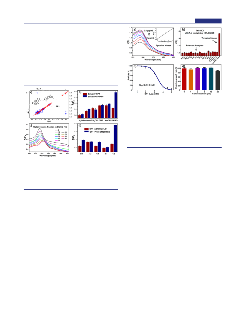
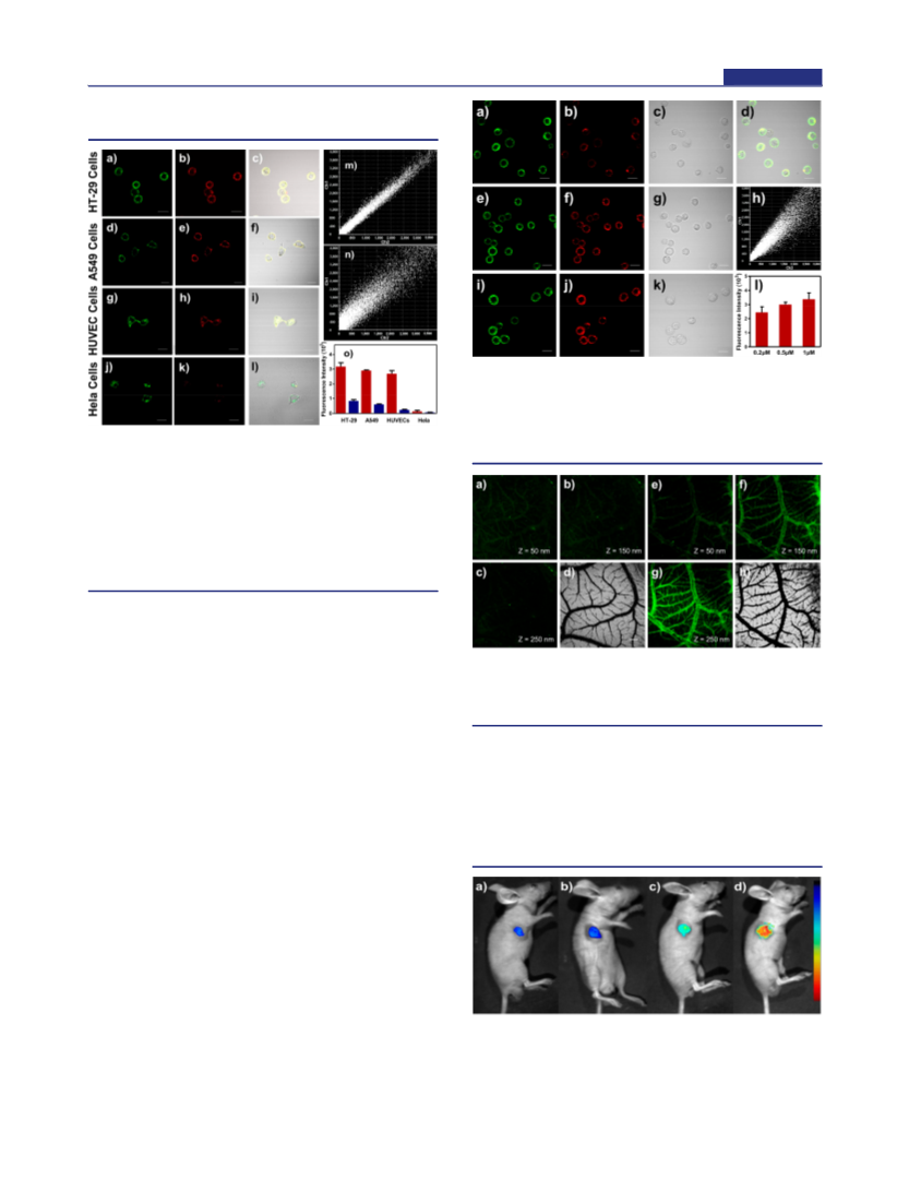
Molecules capable of monitoring receptor protein-tyrosine kinase expression could potentially serve as useful tools for cancer diagnosis due to the overexpression of tyrosine kinases during tumor growth and metastasis. In this work, a conformationally induced "off-on" tyrosine kinase cell membrane fluorescent sensor (SP1) was designed and evaluated for the detection and imaging of receptor protein-tyrosine kinases in vivo and in vitro. SP1 consists of sunitinib and pyrene linked via hexamethylenediamine and displays quenched fluorescence as a dimer. The fluorescence of SP1 is restored in the presence of receptor protein-tyrosine kinases upon strong interaction with SP1 at the target terminal. The unique signal response mechanism enables SP1 use for fluorescence microscopy imaging of receptor protein-tyrosine kinases in the cell membranes of living cells, allowing for the rapid differentiation of cancer cells from normal cells. SP1 can be used to visualize the chick embryo chorioallantoic membrane and mouse model tumors, suggesting its possible application for early cancer diagnosis.

 South Africa | Contact:AAron Thomas
 India | Contact:G.M.
 United States | Contact:sinovainc
 United Kingdom | Contact:Elsa Young
 China (Mainland) | Contact:Xichun Lu