346
S. Yao et al. / Journal of Alloys and Compounds 653 (2015) 345e350
temperature room has been observed when the samples were
synthesized using solidestate reaction method, which is probably
ascribed to the volatility of Bi2O3 and the tendency of Bi3þ to be
oxidized to Bi5þ in the conventional sintering process [15e17]. In
this paper, Ca2Sb2O7:xBi3þ phosphors were prepared by a modified
chemical co-precipitation method, which is one of the most effec-
tive techniques for achieving ultrafine, pure and single-phase
powders and can proceed at moderate temperatures resulting
from enhanced reaction kinetics [18e20]. Finally, we systematically
investigated the photoluminescence properties of the
Ca2Sb2O7:xBi3þ samples and the luminescent mechanism was also
studied.
2. Experimental
2.1. Synthesis of Ca2Sb2O7:xBi nanoparticles
Bi2O3, Sb2O3, CaCl2ꢀ2H2O, NaOH and Na2C2O4 were obtained
from Sinopharm Chemical Reagent Co. Ltd. All the reagents were of
analytical grade and used without further purification. Deionized
water was used throughout the experiment.
Fig. 1. XRD patterns of Ca2Sb2O7:xBi3þ (x ¼ 0, 0.5, 1, 1.5, 2, 4 and 8 mol%) as well as
JCPDS card No. 26e0293 for Ca2Sb2O7. Inset is the amplification of XRD patterns.
The Ca2Sb2O7:xBi precursors were prepared by a chemical co-
precipitation method according to previous work [21]. In brief,
the required amounts of Bi2O3 and Sb2O3 were dissolved in dilute
hydrochloric acid to obtain BiCl3 and SbCl3 solutions, respectively.
Meanwhile, CaCl2 solution was prepared by dissolving CaCl2ꢀ2H2O
in deionized water. Then, BiCl3, SbCl3 and CaCl2 solutions were
mixed together with certain volume ratio according to atom ratio of
Bi:Ca:Sb ¼ x:(2ꢁ1.5x):2 (x ¼ 0, 0.005, 0.01, 0.015, 0.02, 0.04 and
0.08) and formed a clear and homogeneous solution. The mixture
solution was added dropwise into 120 mL of NaOHeNa2C2O4 so-
lution with continuous stirring, and the white precipitate was ob-
tained. The precipitate was washed with dilute ammonia and
anhydrous alcohol for several times, and dried at 80 ꢂC for 10 h.
Finally, the obtained precursor was calcined at 1000ꢂCin air for 4 h.
indexed by weberite structure (JCPDF No. 26e0293). The results
reveal that all the obtained samples are single phase and doping of
Bi3þ at the investigated concentrations does not change the crystal
structure or induce a new phase. However, the diffraction peaks
shift to smaller two theta angles by Bi-doping (inset of Fig.1), which
is in agreement with the qualitative analysis of Bragg's law:
nl ¼ 2dsinq
(1)
In which n,
l, d, and
q
are an integer, wavelength of incident X-
rays (here is 1.5406 Å), distance of lattice planes and Bragg angle,
respectively. When smaller radius of Ca2þ (1.12 nm) is replaced by
large radius of Bi3þ (1.17 nm), the inter-atomic spacing increases
and the lattice expands. As a result, the Bragg angle decreases. This
phenomenon also confirms that Bi3þ ions successfully enter the
crystal lattice of Ca2Sb2O7.
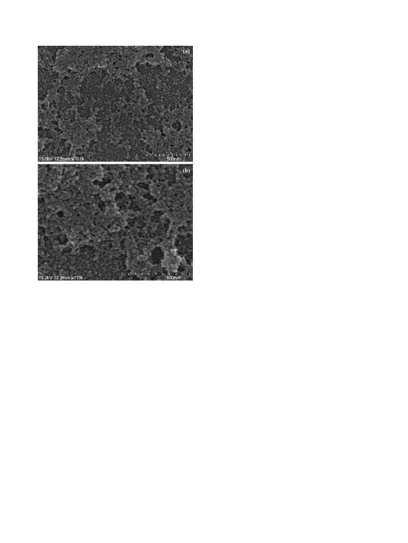
SEM imagines of pure Ca2Sb2O7 and Ca2Sb2O7:0.015Bi samples
are displayed in Fig. 2a and b, respectively. As can be seen, both of
them consist of nearly spherical particles with a particle size in the
range of 20e40 nm. There is little difference in the morphologies
and particle shape between pure Ca2Sb2O7 and Ca2Sb2O7:0.015Bi
samples, indicative of little effect of Bi-doping on the morphology
of Ca2Sb2O7. However, slight aggregation of these particles can be
observed, which might result from the ultrafine effect of the sam-
ples and/or limit of synthetic method.
Fig. 3 shows the XPS spectrum of the Ca2Sb2O7:0.015Bi. From the
whole XPS survey spectrum (Fig. 3a), the Ca2Sb2O7:0.015Bi sample
contains C, Ca, Sb, Bi and O elements, in which the carbon (C 1s:
285 eV) is ascribed to remaining carbonaceous species formed
during the sample preparation and is used for calibration as a
reference. The binding energies of Sb 3d core level in Ca2S-
b2O7:0.015Bi sample are illustrated in Fig. 3b. Two main peaks are
observed at 539.9 eV and 530.5 eV, corresponding to Sb(V) 3d3/2
and 3d5/2 levels, respectively, with a doublet spitting of 9.4 eV,
which matches the typical Sb(V) 3d spineorbit [22]. Furthermore,
the 530.5 eV peak can be fitted into two sub-peaks (Fig. 3c), one
peaking at 530.5 eV and the other centering at 532.1 eV. The latter is
attributed to O 1s binding energy due to the overlapping between
Sb(V) 3d5/2 and O 1s. One should note that no other antimony
species contribution has been found. Fig. 3d gives the high reso-
lution XPS spectra of the Bi 4f region. XPS signals of the Bi 4f can be
found at binding energies of 164.6 eV (4f5/2) and 159.3 eV (4f7/2),
2.2. Characterization
Crystal structure of the Ca2Sb2O7:xBi samples was characterized
by powder X-ray diffractometry on a X'Pert Pro MPD diffractometer
(Pananalytical, Holland) equipped with a Cu Ka radiation source
(l
¼ 1.5406 Å). Morphology of the products was investigated by
scanning electron microscope (SEM, Hitachi S-4600, Japan). The
chemical state identification of the constituent elements was
analyzed by X-ray photoelectron spectroscopy (XPS). XPS spectra
were recorded on an Axis Ultra HAS X-ray photoelectron spec-
trometer using Al Ka radiation with a resolution of 0.68 eV.
The photoluminescence (PL) spectra were obtained on an F-
2500 fluorescence spectrometer (Hitachi, Japan) at room temper-
ature. The diffuse reflectance spectra (DRS) of samples were ob-
tained on a UV-3150 spectrophotometer (Shimadzu, Japan) using
BaSO4 as
a reference standard in the wavelength range of
200e800 nm. The third harmonic (355 nm) of a pulsed Nd:YAG
laser (Spectron Laser Sys. SL802G) was employed to examine the
luminescence decay and decay profiles were recorded with a
LeCloy 9301 digital storage oscilloscope in which the signal was fed
from PMT.
3. Results and discussion
3.1. Characterization
Fig. 1 shows the XRD patterns of the pure and Bi3þ-doped
Ca2Sb2O7 at different concentrations. It can be clearly seen that all
samples have the similar patterns and all diffraction peaks are well

 Yao, Shiyue