Journal of Natural Products
Article
(H11), 5.87 (H12); HR-ESITOFMS m/z 1165.3756 [M + K]+ (calcd
for C57H63F9O13K 1165.3757).
Tris(S)-MTPA ester of 2 (4b). In the same manner as described for
evaporated to dryness under reduced pressure. The residue was
subjected to (R)-PGME amidation and the following LC/MS analysis.
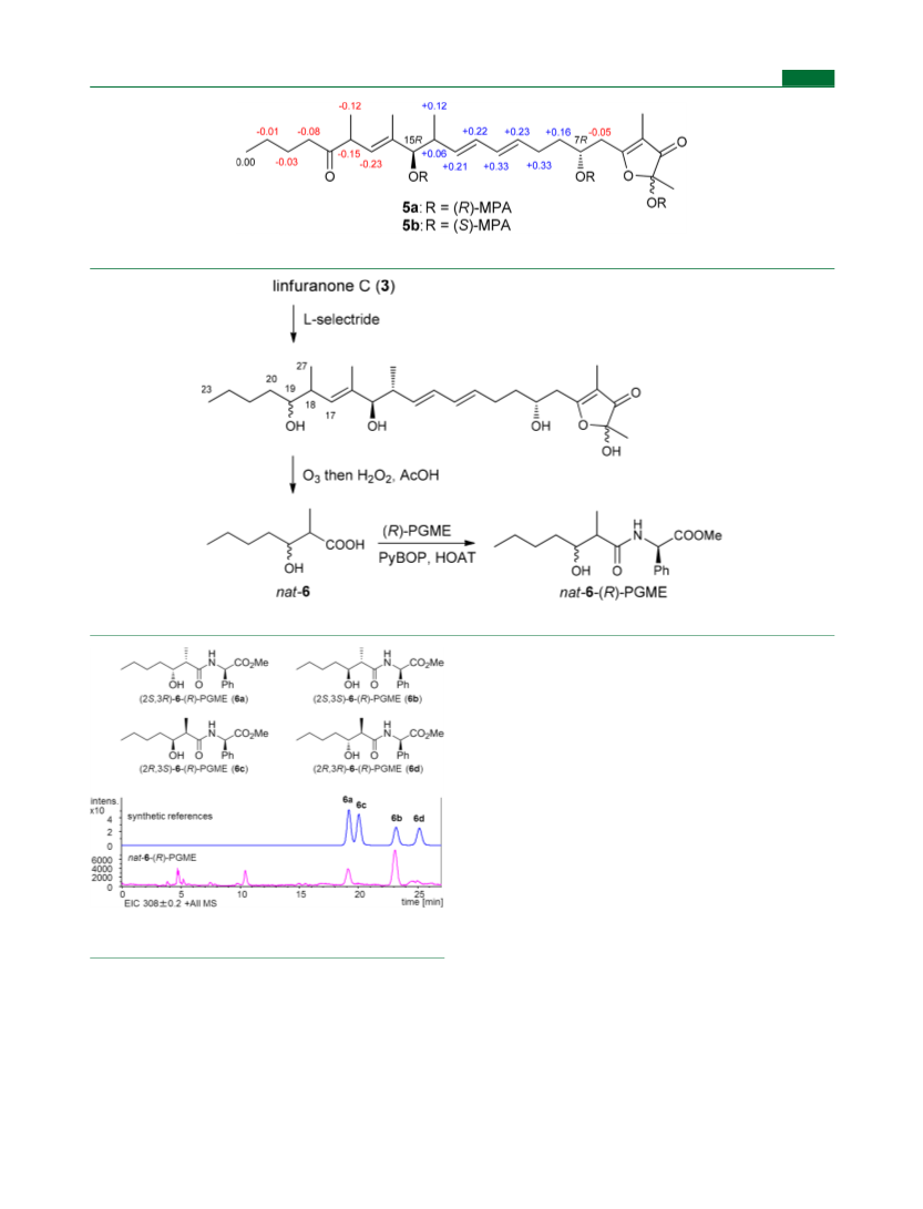
LC/MS analysis of the degradation product of 3. The (R)-PGME
derivative of 3-hydroxy-2-methylheptanoic acid (6) derived from the
degradation of 3 was compared with the (R)-PGME derivatives of
four possible stereoisomers of 6 prepared by chemical synthesis (see
Agilent HP1200 system and Bruker micrOTOF Focus using an ODS
column (COSMOSIL 5C18-AR-II, 2.0 × 150 mm) at a flow rate of 0.3
mL/min with an isocratic elution (40% MeCN in distilled water).
Retention times of the standard samples were 19.3 min for (2S,3R)-6,
20.0 min for (2R,3S)-6, 23.1 min for (2S,3S)-6, and 25.1 min for
(2R,3R)-6. Under the same conditions, the (R)-PGME derivative of
the degraded product of 3 gave two peaks, at 19.1 and 23.1 min,
corresponding to (2S,3R)- and (2S,3S)-isomers, respectively.
Adipocyte Differentiation Assay. The assay was carried out
according to the reported procedure.18 In brief, mouse-derived ST-13
preadipocytes were plated at 1 × 10⁴ cells/mL in 24-well dishes
containing 1 mL of a basal medium containing DME/F-12, 10% fetal
bovine serum, 100 units/mL penicillin, and 100 μg/mL streptomycin
on day 1 and cultured at 37 °C in a humidified atmosphere containing
5% CO2. Moreover, the fresh basal medium containing the indicated
concentrations of linfuranones (or 20 nM rosiglitazone (positive
control)) was replaced on day 2, 5, and 8. On day 12, the ST-13 cells
were washed three times with phosphate-buffered saline, fixed with
10% formalin neutral buffer solution at room temperature for 10 min,
and then washed with distilled water to remove formalin solution.
Furthermore, the cells were rinsed with 60% 2-propanol for 5 min,
stained with 0.24% Oil Red O at room temperature for 20 min, and
then photographed under a phase contrast microscope (100×
magnification) equipped a CCD camera (Leica Microsystems Japan,
Tokyo, Japan).
4a, 4b (2.7 mg, 55% yield) was prepared from 2 (2.0 mg) and (R)-
1
MTPA chloride: H NMR (500 MHz, CDCl3) δ 0.89 (H25), 0.91
(H23), 1.25 (H27), 1.35 (H22), 1.51 (H26), 1.57 (H21), 1.61 (H1),
1.68 (H1), 1.78 (H8), 1.83 (H24), 1.84 (H24), 2.13 (H9), 2.28
(H20), 2.55 (H14), 2.71 (H6), 2.92 (H6), 5.44 (H13), 5.50 (H17),
5.55 (H18), 5.95 (H11), 6.02 (H12); HR-ESITOFMS m/z
1149.4010 [M + Na]+ (calcd for C57H63F9O13Na 1149.4017).
Alkaline hydrolysis of 2 to linfuranone A (1). Compound 2 (5.0
mg, 10 μmol) was dissolved in 5% NaOMe/MeOH (2.6 mL) and
stirred for 30 min at room temperature. The reaction mixture was
neutralized by adding 5% HCO2H solution (4 mL) and then
evaporated to dryness. The crude material was purified by HPLC
using a Cosmosil C18-AR-II column (10 × 250 mm) with an isocratic
solvent system of 30% MeCN in distilled water at a flow rate of 4.0
mL/min, monitoring at 280 nm to yield 1 (2.3 mg, tR = 12.1 min):
1
[α]23 −3.0 (c 0.10, MeOH); H and 13C NMR data matched those
D
of naturally occurring linfuranone A (1); HR-ESITOFMS m/z
393.2285 [M − H]− (calcd for C22H33O6 393.2283).
Oxidative Degradation of 2 and Chiral HPLC Analysis. To a
solution of 2 (2.0 mg, 4.2 μmol) in EtOAc (5 mL) was bubbled O3 at
−78 °C until the solution turned pale blue. After bubbling oxygen
into the solution at the same temperature for 15 min, HCO2H (0.75
mL) and 30% H2O2 (0.44 mL) were added, and the reaction mixture
was heated at 60 °C for 15 h. Then, the reaction mixture was
evaporated under vacuum, the resultant material was dissolved in
MeOH (200 μL), and 28% NaOMe solution (150 μL) was added.
After standing for 18 h at ambient temperature, the reaction mixture
was analyzed on a chiral HPLC (Sumichiral OA-5000, 4.6 × 150 mm;
1 mM CuSO4 solution; 1.0 mL/min; detection at 254 nm). Retention
times of the standard samples were 21.8 min for (S)-lactic acid and
27.8 min for (R)-lactic acid. Co-injection of the degraded product of 2
gave two peaks when injected with (S)-lactic acid and a single peak
when injected with (R)-lactic acid.
Tris(R)-MPA ester of 3 (5a). To a solution of 3 (1.0 mg, 2.2 μmol)
in dry CH2Cl2 (120 μL) were added (R)-MPA (1.5 mg, 9.0 μmol),
N,N-dimethyl-4-aminopyridine (DMAP, trace amount), and a
solution of diisopropylcarbodiimide (DIC, 1 mg, 7.9 μmol) in dry
CH2Cl2 (20 μL) at room temperature. After standing for 30 min, the
reaction mixture was diluted with ice/water and extracted with
EtOAc. The organic layer was concentrated under reduced pressure,
and the residue was purified by HPLC (n-hexane/EtOAc, 8:1−1:1) to
give tris(R)-MPA ester 5a (0.6 mg, 30% yield): 1H NMR (500 MHz,
CDCl3) δ 0.88 (H23), 0.90 (H25), 0.93 (H27), 1.22 (H22), 1.43
(H21), 1.70 (H8), 1.98 (H9), 2.22 (H20), 2.49 (H14), 2.72 (H6),
3.16 (H18), 5.06 (H17), 5.40 (H13), 5.47 (H10), 5.85 (H11), 5.96
(H12); HR-ESITOFMS m/z 929.4445 [M + Na]+ (calcd for
C54H66O12Na 929.4446).
Tris(S)-MPA ester of 3 (5b). In the same manner as described for
5a, 5b (0.5 mg, 25% yield) was prepared from 3 (1.0 mg, 2.2 μmol)
and (S)-MPA: 1H NMR (500 MHz, CDCl3) δ 0.78 (H25), 0.88
(H23), 1.05 (H27), 1.23 (H22), 1.46 (H21), 1.54 (H8), 1.66 (H9),
2.29 (H20), 2.44 (H14), 2.76 (H6), 3.31 (H18), 5.19 (H13), 5.23
(H10), 5.29 (H17), 5.52 (H11), 5.74 (H12); HR-ESITOFMS m/z
929.4447 [M + Na]+ (calcd for C54H66O12Na 929.4446).
Degradation of 3. To a stirred solution of 3 (0.25 mg, 0.5 μmol)
in dry THF (50 μL) was added L-selectride (1.0 M THF solution, 10
μL, 0.5 μmol) at 0−5 °C. After 1 h, L-selectride (40 μL, 0.5 μmol) was
further added to the solution, which was then stirred for 18 h at
ambient temperature. The reaction was quenched with a saturated
NH4Cl solution and extracted with EtOAc. The organic layer was
dried over anhydrous Na2SO4 and concentrated under reduced
pressure. The residual material was dissolved in EtOAc (5 mL), to
which was bubbled O3 at −78 °C for 30 min. Then, formic acid (0.75
mL) and 30% H2O2 solution (0.40 mL) were successively added to
the reaction mixture, and the temperature was gradually raised to 60
°C. After stirring for 15 h at the same temperature, the mixture was
ASSOCIATED CONTENT
* Supporting Information
■
S
The Supporting Information is available free of charge on the
Experimental procedures and 1D and 2D NMR spectra
of compounds 2 and 3 (PDF)
AUTHOR INFORMATION
Corresponding Author
*Tel: +81-766-56-7500. Fax: +81-766-56-2498. E-mail: yas@
■
ORCID
Notes
The authors declare no competing financial interest.
ACKNOWLEDGMENTS
■
This work was supported by JSPS KAKENHI Grant Number
24580156 to Y.I., Core-to Core Program, JSPS, and National
Research Council of Thailand (NRCT) to A.T.
REFERENCES
■
(1) (a) Van Lanen, S. G.; Shen, B. Curr. Opin. Drug Dev. 2008, 11,
186−195. (b) Chen, H.; Du, L. Appl. Microbiol. Biotechnol. 2016, 100,
541−557.
(2) (a) Capon, R. J.; Faukner, D. J. J. Org. Chem. 1984, 49, 2506−
2508. (b) Cimino, G.; Sodano, G.; Spinella, A. J. Org. Chem. 1987, 52,
5326−5331. (c) Kuroda, K.; Yoshida, M.; Uosaki, Y.; Ando, K.;
Kawamoto, I.; Oichi, E.; Onuma, H.; Yamada, K.; Matsuda, Y. J.
Antibiot. 1993, 46, 1196−1202. (d) Paul, M. C.; Zubia, E.; Ortega, M.
J.; Salvia, J. Tetrahedron 1997, 53, 2303−2308. (e) Kunze, B.;
H
J. Nat. Prod. XXXX, XXX, XXX−XXX

 Akiyama, Hirofumi