K. Anupriya et al. / Journal of Alloys and Compounds 590 (2014) 406–410
407
2
. Experimental
2.1. Materials required
Chemicals used in this synthesis were Cerium (III) Nitrate hexa hydrate (purity
of 99.99%,Ce(NO ) as a precursor, CTAB (cetyltrimethylammonium bromide) as
3 2
)
surfactant and Monoethanolamine as a reducing agent, all chemicals being pur-
chased from CDH chemicals and Merck TM. Ethylene glycol (>99.99% pure, Merck
TM) was used as a solvent Conc. HCl was used to adjust the pH. All these chemicals
were used without any further purification.
2.2. Synthesis
In a typical synthesis, 0.2 M of Cerium Nitrate and 0.2 g of CTAB were mixed in
ethylene glycol (EG)/water (1:1) mixed solvent system. The reaction was performed
at room temperature at a constant stirring rate. The initial pH was noted to be 7. Few
drops of concentrated HCl were added to the mixture to reduce the pH to 1. Mono-
ethanolamine was then dripped to the above solution until the pH got increased to
1
3
2. Initially a yellow solution of cerium hydroxide Ce(OH) was obtained. Slowly, this
yellow color disappears and turns to purple. This is due to absorption of dissolved
oxygen from air which is an intermediate mixed hydrate state. Stirring was carried
out in an ambient environment for 12 h. Finally, yellow precipitate was obtained
showing the complete conversion of cerium oxide nanoparticles. Then, the solution
was centrifuged at 7500 rpm and washed with acetone and double distilled water
for three times. It was then dried at 100 °C for 2 h in hot air oven. Further the dried
powder was calcinated at 400 °C for 2 h to obtain a crystalline product.
2.3. Materials characterization
Powder XRD analysis was carried out using a Bruker AXS D8 Advance X-ray dif-
fractometer using Cu Ka radiation (k = 0.15406 nm) with a scintillation counter. The
ꢀ
1
scanning rate was 0.05 s in the 2h range from 20° to 100°. The specific surface
area was measured with Quantachrome Nova Win 3200e equipment by de-gassing
the sample at 200 °C for 2 h and specific area was calculated by nitrogen adsorption
keeping the relative pressure between 0.04 and 0.2. TEM studies were performed
with a JEOL JEM-200CX instrument operating at 200 kV. EDS analysis was per-
formed with a HITACHI S-3700N scanning electron microscope with an accelerating
voltage of 20 kV. Surface morphology was examined using a XE-100 Park systems
AFM, operated in a non-contact mode with a scan rate of 1 Hz. Scan size in X and
Fig. 1. XRD pattern of cerium oxide nanoparticles prepared by chemical precipi-
tation route. (a) Reference JCPDS card 43–1002, (b) before calcination of ceria NP’s,
Y axis was made as 1 ꢁ 1
lm. Raman spectra were collected using a Renishaw Invia
(
c) after calcinated at 400 °C.
Raman microscopy at room temperature. He–Ne laser beam with an excitation of
6
1
33 nm was focused under 50ꢁ objective lens and the laser spot size was about
ꢀ1
l
m. Raman spectra were recorded in the 200–600 cm range with the spectral
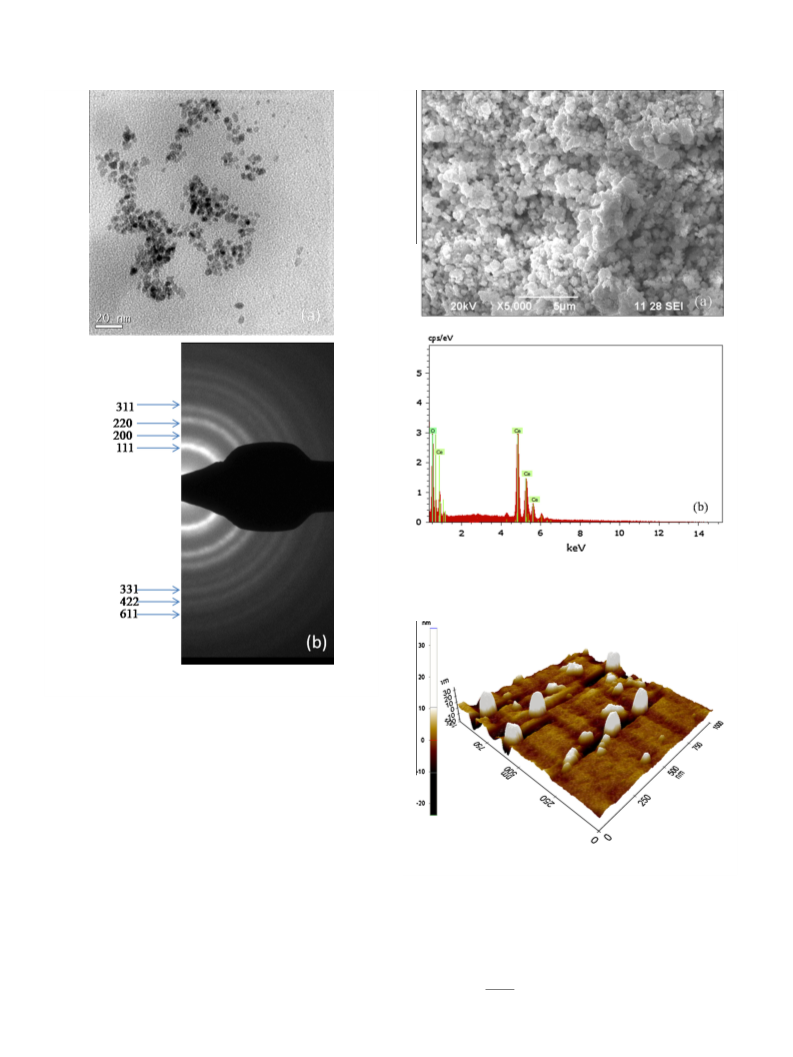
pattern as in Fig. 2(b). Like the XRD profile, the reflections were in-
dexed to 111, 200, 220, 311, 331, 422, 611 planes respectively with
ꢀ
1
resolution of 1 cm . The UV–Visible spectra were recorded using V-650 UV–Vis
instrument from JASCO operated at room temperature in the wavelength between
2
00–700 nm with a scan speed of 400 nm/min.
2
a cubic CeO crystal structure. In addition, all the diffraction rings
match with CeO
system.
The specific surface area is about 139 m /g exhibited for cerium
oxide nanoparticles measured by nitrogen adsorption with a rela-
tive pressure of 0.04–0.2. The corresponding particle size (dBET) can
be estimated as [11]
2
powder showing absence of impurity in the
3
. Results and discussion
2
3.1. Structural analysis
The X-ray diffraction (XRD) pattern obtained from as-prepared
and calcinated ceria is as shown in Fig. 1, the improvement in crys-
tallinity after calcination. These peaks are in good agreement with
the standard JCPDS card 43–1002 for face centered cubic structure.
Before calcination as in Fig. 1b shows poor crystallinity with weak
d
BET ¼ 6=
q
A
ð1Þ
3
where
q is the true density of CeO , i.e. 7.28 g/cm and A is the spe-
2
cific surface area calculated from BET. The particle size estimated
from BET indicates that the average crystalline size is about
5
peaks viewing the presence of Ce(OH)
was calcinated at 400 °C [26] where Ce(OH)
rectly to form CeO . Thus, the effect of calcination temperature
4
. So the resultant sample
2
.9 nm for CeO nanoparticles.
4
will be dehydrated di-
2
shows a good crystalline growth of cerium oxide NP’s with a crys-
talline sharp and strong diffraction peak. The diffraction peaks
were all relatively broader because of the smaller size of cerium
oxide nanoparticles. The crystallite size was about 9 nm calculated
by using the Debye–Schrrer’s formula.
From the Fig. 1c, the characteristic peaks of cerium oxide nano-
particles after calcination at 400 °C are in consistent with face cen-
tered cubic structure.
The average particle size of cerium oxide nanoparticles was
examined by using TEM. The results confirmed that the particles
were in spherical shape and also the size distribution was uniform
with an average crystallite size of 4 nm as in Fig. 2(a). The highly
3.2. Morphological studies
Structural morphological studies were done using SEM as
shown in Fig. 3(a). It shows that nanostructured CeO was nearly
2
spherical in shape with a variety of particle sizes due to the calci-
nation which makes the particles condense faster. Aggregates seen
above are due to the accumulation of nanoparticles. It can be seen
that the average diameter of the secondary particles were ꢂ36 nm
to ꢂ50 nm respectively, along with fine particles below ꢂ10 nm. As
crystallite size was not clearly identified by using SEM further
investigation was done by using TEM. The energy dispersive X-
ray (EDAX) spectrum gives information about the presence of Ce
and O. This confirms that the resultant sample was cerium oxide
and is shown in Fig. 3(b).
2
crystalline nature of CeO powder leads to the Debye–Scherrer
diffraction rings in the Selected Area Electron Diffraction (SAED)

 Anupriya