Z.-J. Luan et al. / Journal of Molecular Catalysis B: Enzymatic 116 (2015) 78–82
79
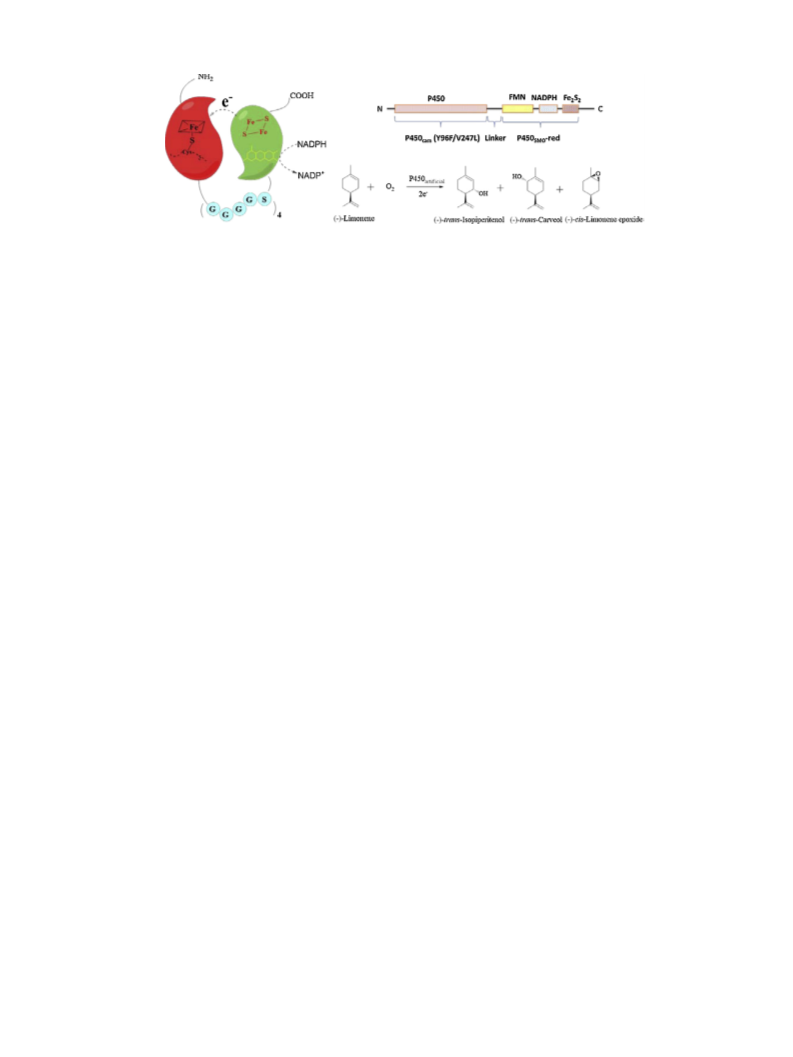
Scheme 1. Construction of an artificial P450 system: P450cam (Y96F/V247L)–P450SMO red.
Class I P450. So we speculated whether the P450SMO reductase
domain could act as an effective redox partner for generating effi-
cient P450 chimeras.
(pH 7.5), and disrupted by sonification. After centrifugation, the
soluble fractions were subjected to spectrophotometric analysis or
reductase domain activity assay.
Cytochrome P450cam (PdR, Pdx) catalyzes the oxidation of the
bicyclic compound d-(+)-camphor to 5-exo-hydroxycamphor, the
first step in the camphor metabolism pathway of the soil bacterium
Pseudomonas putida. The Y96F/V247L mutant of P450cam could cat-
alyze the hydroxylation of (−)-limonene in one step, producing
2
.3. Spectrophotometric analysis and reductase activity assay of
P450a F1–F4
To quantitate the content of active cytochrome P450 in the
(
−)-trans-isopiperitenol, (−)-trans-carveol and (−)-cis-limonene
recombinant E. coli, the soluble fraction of P450a F1–F4 were used
to measure the CO-reduced P450 absorption with a UV-vis spec-
trophotometer [19]. The reductase activity was determined by
measuring the increase in absorbance at 550 nm due to the reduc-
tion of cytochrome c [20].
epoxide [18]. In the present work, we therefore constructed a
self-sufficient chimera P450cam (Y96F/V247L)–P450SMO red, by fus-
ing the P450SMO reductase domain with the P450cam mutant
domain together (see Scheme 1). By optimizing the linker sequence
between the two domains and co-expressing with glucose dehy-
drogenase (GDH, used for NADPH regeneration), the whole cell
could be used for monoterpene hydroxylation, indicating that the
P450SMO reductase might be used as the electron transfer partner
for heterologous P450s. Furthermore, the biosynthetic P450s (Class
I) lacking such a universal reductase may be engineered similarly
into diverse self-sufficient P450s for either functional characteri-
zation or potential application.
2
.4. Bioconversion of (−)-limonene by the fused enzymes with
different linkers
To determine the catalytic activity of the fused enzymes with
different linkers, we performed a 1 mL reaction with the cell free
extracts. The reaction mixtures containing 50 mM potassium phos-
phate buffer (pH 7.5), 1 M P450a F1–F4, 100 M (−)-limonene and
◦
1
00 M NADPH were incubated at 25 C. After incubation for 12 h,
2
. Experimental
the reaction mixture was extracted with 900 L ethyl acetate by
vigorous shaking. The organic layer was separated by centrifuga-
tion, dried over anhydrous sodium sulfate, and then subjected to
GC–MS analysis.
2.1. Engineering the chimeric P450cam (Y96F/V247L)–P450SMO red
F1–F4
The negative control was conducted under the same condition
using the same amount P450cam (Y96F/V247L) and the P450SMO
reductase, which were not fused together. The positive control was
conducted by P450cam system under the same condition, which
contained P450cam (Y96F/V247L), putidaredoxin reductase (PdR)
and putidaredoxin (Pdx).
The genes of P450cam and P450SMO were cloned from P. putida
PpGl (ATCC17453) and Rhodococcus sp. ECU0066, respectively. Site-
directed mutagenesis of P450cam (Y96F/V247L) gene was carried
out with the Quick Change® Site-directed Mutagenesis Kit. The
primers for the mutagenesis were shown in Table S1 (Supporting
Information).
As shown in Table S1, P450cam (Y96F/V247L) and P450SMO
reductase were amplified with primers A, C1/D1, C2/D2, C3/D3,
C4/D4, B to construct four different linkers. The amplified P450 gene
2.5. Plasmid construction for co-expression of P450cam
(Y96F/V247L)–P450SMO red F3 and GDH genes
ꢀ
ꢀ
has NdeI site at 5 -ends and HindIII at the 3 -ends. Meanwhile, the
ꢀ
ꢀ
P450SMO red gene has HindIII site at 5 -ends and XhoI at the 3 -ends.
Then the two genes were ligated into pET28a(+) and fused together.
The fused enzymes P450cam (Y96F/V247L)–P450SMO red F1–F4 have
the His-tag at the N-terminal for subsequent purification. The fused
enzymes are abbreviated as P450a F1–F4 in this article.
Plasmids and the primers used for co-expression in this study
are listed in Tables S2 and S3. For construction of the co-expression
system, the glucose dehydrogenase (GDH) gene was amplified with
primers 1 and 2 using genome DNA from Bacillus megaterium. The
resulting 786 bp fragment was digested with NdeI and XhoI and
then ligated into pACYCDuet-1 which was digested with the same
restriction enzymes, generating the plasmid pACYCDuet-1-GDH.
Successful ligation into pACYCDuet-1 was confirmed by restriction
analysis.
2.2. The expression of fusion enzymes
Recombinants containing pET28a(+)-P450a F1–F4 were grown
in 100 mL Luria-Bertani (LB) medium with 100 g/mL of kanamycin
at 37 C. After the optical density OD600 had reached 0.5, 0.2 mM
Co-expression of P450a F3 and GDH genes was performed
in a two-plasmid system with different origins for replication,
P450a and GDH genes were respectively cloned into pET28a(+)
and pACYCDuet-1. The same E. coli strain transformed with both
pET28a(+)-P450a and pACYCDuet-1-GDH was named as BL21-
pET28a-P450a F3/pACYCDuet-GDH (see Scheme 2).
◦
isopropyl-ˇ-d-thiogalactopyranoside (IPTG) and 0.2 mM ı-amino
levulinic acid (ı-ALA) were added, and then the cultivation was
◦
continued with reciprocal shaking for 18 h at 25 C. The cells
were collected, suspended in 50 mM potassium phosphate buffer

 Luan, Zheng-Jiao