D-Pro (6.5) in solvent B. The retention times and ESIMS
product ions (m/z [M+Na] ) of the L-FDAA derivatives of
N-Me-Ile and N-Me-Val from the hydrolyzate were 6.6 min
J = 6.9 Hz, 3H), 0.99 (m, 1H), 0.95 (d, J = 6.5 Hz, 3H), 0.91 (d,
+
J = 6.8 Hz, 3H), 0.85 (d, J = 6.7 Hz, 3H), 0.84 (t, J = 7.4 Hz,
+
3H); HRESIMS m/z [M+Na]
727.2180 (calcd for
(
420.1) and 5.3 min (406.1) in solvent A, respectively, proving
C H BrF N O Na, 727.2176).
3
2
44
3
2
7
1
the configurations of N-Me-Ile and N-Me-Val were L. The
retention times and ESIMS product ions (m/z [M+Na] ) of the
L-FDAA derivatives of Val and Pro from the hydrolyzate were
6
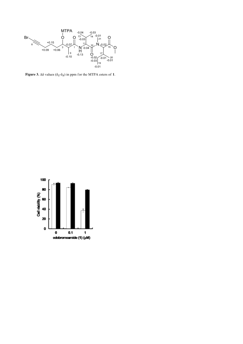
R-MTPA ester: H NMR (500 MHz, CDCl ) δ 6.29 (d, J =
3
+
8.7 Hz, 1H), 5.28 (q, J = 6.0 Hz, 1H), 5.03 (d, J = 10.4 Hz, 1H),
4.74 (dd, J = 8.6, 7.0 Hz, 1H), 3.69 (s, 3H), 3.07 (s, 3H), 2.54
(quin, J = 6.6 Hz, 1H), 2.08 (td, J = 7.1, 2,6 Hz, 2H), 2.01 (m,
1H), 1.98 (m, 1H), 1.64 (m, 2H), 1.35 (m, 1H), 1.32 (m, 2H),
1.19 (d, J = 7.0 Hz, 3H), 1.02 (m, 1H), 0.96 (d, J = 6.7 Hz, 3H),
0.94 (d, J = 6.9 Hz, 3H), 0.89 (d, J = 6.8 Hz, 3H), 0.85 (t, J =
.8 min (392.1) and 4.9 min (390.1) in solvent B, respectively,
proving the configurations of Val and Pro were L.
Absolute Configuration of Hmba Unit of 1.
The
absolute configuration of the Hmba unit in 1 was determined
by derivatization with HPLC labeling reagent and chiral HPLC
analyses of the derivatives. Hmba from the acid hydrolyzate of
+
7.4 Hz, 3H); HRESIMS m/z [M+Na] 727.2170 (calcd for
C H BrF N O Na, 727.2176).
3
2
44
3
2
7
1
was treated with 80 μL of a phenacyl bromide solution (12
Brine Shrimp Toxicity Assay.
The brine shrimp
mg/mL in acetone) and 80 μL of a triethylamine solution (10
mg/mL in acetone), and the mixture was stirred at 50 °C for 6 h.
The reaction mixture was subjected to chiral HPLC [DAICEL
CHIRALPAK IC (4.6×250 mm), n-hexane/EtOH=90:10 at 1.0
mL/min, UV detection at 254 nm]. The phenacyl esters of
authentic standards were prepared by the same procedure. The
retention times (min) of the authentic standards were L-Hmba
(Altemia salina) toxicity assay was performed using a slight
modification of the original method. The samples were
dissolved in MeOH and transferred to 0.7 cm squares of filter
paper. The squares were dried and added to test vials of
artificial seawater (2.0 mL). Approximately 10 hatched brine
shrimp were transferred to the vials. After 24 h at room
temperature, the brine shrimp were counted and the percentage
of live verses total shrimp was calculated to determine the LD50
values.
(
12.2 min) and D-Hmba (21.2 min). The retention time and
+
ESIMS product ion (m/z [M+Na] ) of Hmba in 1 was 12.2 min
(
259.1), proving the configuration of Hmba was L.
MTT Assay.
The cytotoxicity against human cell
Isolation of Hmoaa Moiety. A solution of 1 (5.4 mg) in
lines HeLa S3 was measured by means of the MTT assay. HeLa
S3 cells were seeded at 4×10 cells/well in 96-well plates and
3
MeOH (1.0 mL) was treated with 10% Pd/C catalyst and stirred
under an atmosphere of hydrogen at room temperature for 24 h.
The reaction mixture was filtered, concentrated in vacuo and
subjected to HPLC [COSMOSIL 5C -AR-II (10×250 mm),
cultured overnight at 37 °C with 5% CO2. Various
concentrations of compound were added and the culture plates
were kept for 72 h. MTT solution (20 μL, 5 mg/mL in DMSO)
was added to each well and the plate was further incubated for
4 h. After the incubation, all remaining supernatant were
removed and DMSO (150 mL) was added to each well to
dissolve the resultant formazan crystal. Absorbance was
measured by using a microplate reader at a wavelength of 540
nm, using 630 nm as the reference wavelength. The IC50 values
were calculated by curve fitting method.
1
8
8
5% MeOH at 4.0 mL/min, UV detection at 215 nm] to yield
the hydrogenation product of 1. The product was treated with
M HCl (0.5 mL) at 105 °C for 24 h. The hydrolysate was
3
concentrated to dryness and partitioned between H O and
2
EtOAc. The EtOAc layer was separated by column
chromatography on silica gel using n-hexane/EtOAc (3:1 to
0
:1) to give Hmoaa.
1
Hmoaa: H NMR (500 MHz, CDCl ) δ 3.95 (m, 1H), 2.63
Trypan Blue Dye Exclusion Assay.
HeLa S3 cells
3
4
(
dq, J = 7.2, 3.6 Hz, 1H), 1.54–1.29 (m, 8H), 1.22 (d, J = 7.2
were seeded at 8×10 cells/well in 24-well plates, cultured
overnight and then preincubated with or without 50 μM
Z-VAD-FMK for 30 min. The cells were then treated with
various concentrations of compound for 48 h. They were then
stained with trypan blue, and the number of stained cells was
counted.
+
Hz, 3H), 0.90 (t, J = 6.9 Hz, 3H); HRESIMS m/z [M+Na]
97.1126 (calcd for C H O Na, 197.1148).
1
9
18
3
Preparation of MTPA Esters.
A solution of 1
(
6.9 mg) in 5% methanolic KOH (0.5 mL) was stirred for 24 h
at room temperature. The reaction mixture was concentrated,
and the residue was partitioned between CH Cl and H O. The
2
2
2
organic layer was subjected to reversed-phase HPLC [Cosmosil
C -AR-II (10×250 mm), 65% MeOH with 0.1% TFA at 4.0
We thank Dr. T. Yamori (Screening Committee of
Anticancer Drugs supported by a Grant-in-Aid for Scientific
Research on Priority Area “Cancer” from The Ministry of
Education, Culture, Sports, Science and Technology, Japan) for
screening in the human cancer cell line panel. This work was
supported in part by a Grant-in-Aid for Young Scientists (B)
(21710237) from the Ministry of Education, Culture, Sports,
Science and Technology, Japan; University of the Ryukyus
Strategic Research Grant.
5
1
8
mL/min, UV detection at 215 nm] to yield compound 2. The
product was treated with trimethylsilyldiazomethane (10 μL) in
benzene/MeOH (2:1, 60 μL), and the mixture was stirred for 2
h at room temperature, to give methyl ester 3. This compound
was divided into two equal portions (0.3 mg each), and one
portion was reacted with R-MTPACl (10 μL) and DMAP (0.5
mg) in pyridine (50 μL), and the mixture was stirred for 20 h at
room temperature. The reaction mixture was concentrated, and
the residue was partitioned between EtOAc/0.1M NaHCO3
Supporting Information
1D and 2D NMR spectra and HCC panel data for
odobromoamide, and gene screening. This material is available
electronically on J-STAGE.
(1:1). The extract obtained from the organic layer was
subjected to reversed-phase HPLC [Cosmosil 5C -AR-II
1
8
(10×250 mm), 85% MeOH at 4.0 mL/min, UV detection at 215
nm] to yield S-MTPA ester. Using the same procedure as
described above, R-MTPA ester was obtained from the other
References
portion of 3.
1.
2.
L. T. Tan, Phytochemistry. 2007, 68, 954.
J. K. Nunnery, E. Mevers, W. H. Gerwick, Curr. Opin.
Biotechnol. 2010, 21, 787.
M. Nagarajan, V. Maruthanayagam, M. Sundararaman, J.
Appl. Toxicol. 2012, 32, 153.
1
S-MTPA ester: H NMR (500 MHz, CDCl ) δ 6.16 (d, J =
3
8
4
.4 Hz, 1H), 5.29 (q, J = 6.2 Hz, 1H), 5.01 (d, J = 10.5 Hz, 1H),
.70 (dd, J = 8.5, 7.2 Hz, 1H), 3.69 (s, 3H), 3.06 (s, 3H), 2.47
3.
4.
(
1
quin, J = 7.0 Hz, 1H), 2.17 (t, J = 7.0 Hz, 2H), 2.00 (m, 1H),
.95 (m, 1H), 1.70 (m, 2H), 1.47 (m, 2H), 1.33 (m, 1H), 1.09 (d,
R. K. Singh, S. H. Tiwari, A. K. Rai, T. M. Mohapatra, J.

 Sueyoshi, Kosuke