D. Xiong et al. / Journal of Alloys and Compounds 642 (2015) 104–110
105
for the synthesis of silver based delafossite oxides are much fewer.
The inherent cause is suggested to derive from the fact that, the
simple binary oxide of silver (Ag2O) is easy to decompose at a tem-
perature of ꢀ300 °C, which limits the preparation of silver based
delafossites through solid-state reactions. Therefore, it becomes a
reasonable choice to synthesize silver based delafossite oxides at
low-temperature and/or closed reaction systems [2,27]. For exam-
After reaching a homogeneous state, the solution was loaded into
a 100 ml Teflon-lined autoclave, which was sealed and maintained
at 190–230 °C for 60 h. After the reaction finished, the obtained
yellow green precipitate was washed with diluted nitric acid,
deionized water and absolute alcohol in sequence for several
times, and then stored in absolute alcohol solution. Finally, about
2.0 g nano-sized AgCrO2 product could be gained from each
reaction.
ple, AgCrO2 crystals with an average size of around 1 lm have been
synthesized through cation exchange reactions, by heating LiCrO2,
KNO3 and AgNO3 at 350 °C in an evacuated silica tube for 4 days
[19,21]. Recently, AgCrO2 crystals with an average size of 2 lm
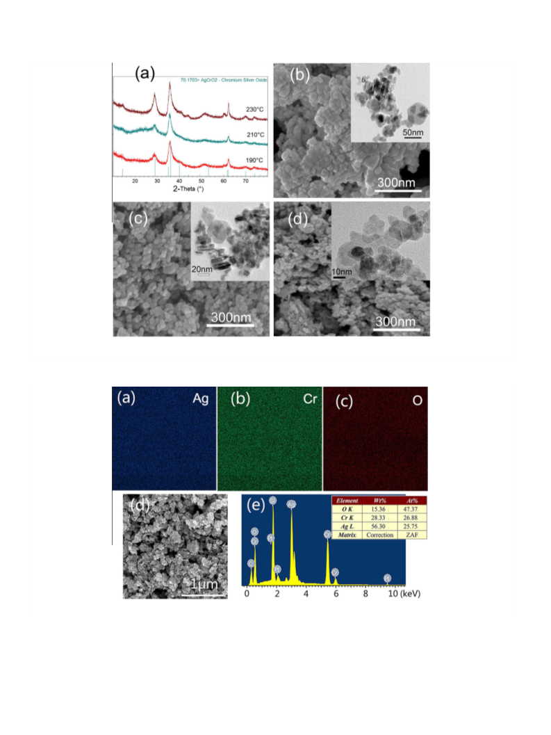
Fig. 1 shows the XRD patterns and morphologies of AgCrO2
nanocrystals prepared at different reaction temperature (190 °C,
210 °C and 230 °C). Fig. 1a shows that all of the diffraction peaks
can be indexed to delafossite AgCrO2 (JCPDS File Card No. 70-
1703) with the hexagonal R3m crystal structure, and no impurity
phase can be detected. The full width at half maximum of the
XRD patterns becomes wider as the reaction temperature
decreases, which reflects the crystal size decreases. By applying
the Scherrer equation to the broadened diffraction peaks, the aver-
age crystal sizes at different synthesis temperatures of 190 °C,
210 °C and 230 °C were calculated to be 18.4 nm, 15.9 nm and
14.1 nm, respectively. This result is consistent with their corre-
sponding SEM and TEM images in Fig. 1b–d. From TEM images,
the nanocrystals are with the hexagonal nanoplates morphology.
The observed diameters of the nanoplates obtained at 230 °C,
210 °C and 190 °C are 15–50 nm, 15–30 nm and 10–30 nm, respec-
tively, while their thicknesses are much thinner. From these, it is
known that the decreased reaction temperature leads to slower
crystal growth rate, and therefore smaller crystal size of the
nano-products.
have been prepared under a critical hydrothermal condition
(400 °C, 40 MPa) [27]. Note that, those reported processes are com-
plicated and include tough conditions; to synthesize nano-sized
silver based delafossite oxides still remains as great challenge.
In this work, delafossite AgCrO2 nanocrystals with ultrafine
sizes of 10–20 nm have been synthesized for the first time via a
low temperature hydrothermal method, and its potential as photo-
cathode in p-type DSSC has been primarily examined. The synthe-
sis parameters effecting on the crystal phases and morphologies
have been studied. The element composition and valence state
information, thermal stability, electrical and optical properties of
AgCrO2 nanocrystals have been systematically investigated. At last,
a general crystal formation mechanism for the hydrothermal syn-
thesis of chromium based delafossite oxides (AgCrO2 and CuCrO2
nanocrystals) based on the in-situ oxidation–reduction reactions
have been proposed.
Furthermore, the SEM–EDS mapping results of AgCrO2
nanocrystals are shown in Fig. 2, to reduce the measurement errors
caused by absorption of water molecules on the sample surface,
the AgCrO2 was analyzed after annealing at 120 °C for 2 h in vac-
uum. It can be observed that all of the Ag, Cr and O elements are
homogeneously distributed (Fig. 2a–c), the elementary percent-
ages of Ag (25.75 at.%), Cr (26.88 at.%), O (47.37 at.%) are nearly
consistent with their source materials’ concentrations in the
hydrothermal precursor and close to the stoichiometric proportion
of AgCrO2 (Ag: Cr: O = 1: 1.04: 1.84, see Fig. 2e). In addition, the
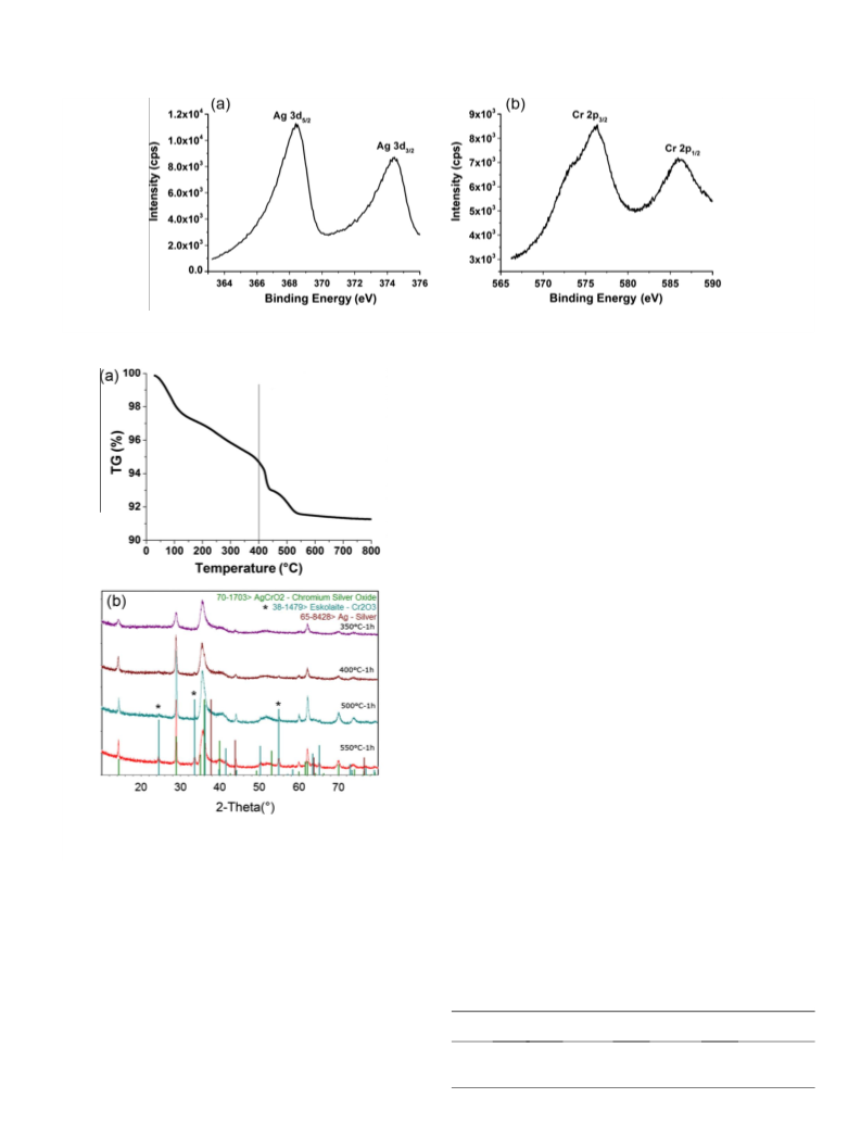
elemental chemical states of the AgCrO2 crystals have been inves-
tigated by XPS. The corresponding results are shown in Fig. 3. The
peaks located at near 368.5 eV (Ag 3d 5/2) and 373.9 eV (Ag 3d 3/2)
shown in Fig. 3a confirm the monovalent state of silver element
(Ag+) in the samples [20]. The peaks located at near 576.3 eV and
585.8 eV (Fig. 3b) are corresponding to the binding energies of Cr
2p 3/2 and Cr 2p 1/2, which confirm the trivalent state of chro-
mium element (Cr3+) in the samples [12,26].
2. Experimental section
All of the chemicals in these experiments without special notification were pur-
chased from Sigma Aldrich with analytical grade and used without further purifica-
tion. In
a typical hydrothermal synthesis, certain amounts of reactants were
dissolved in deionized water, and the obtained solution was transferred into a
Teflon-lined autoclave. The sealed autoclave was maintained at 190–230 °C for
reaction. After the reaction finished, the autoclaves were naturally cooled to room
temperature. Finally, the obtained precipitate was washed for several times in a
centrifugal cleaning machine and was finally stored in absolute alcohol solution
for further use.
Powder X-ray diffraction patterns were collected at room temperature by using
a Panalytical X’pert Pro diffractometer (XRD, Cu K
a radiation). A field emission
scanning electron microscope (FESEM) system (Hitachi-S-4800) coupled with
energy dispersive X-ray spectroscopy (EDX) and a transmission electron micro-
scope (FETEM, Tecnai G2 F30) were used to observe the microstructure and deter-
mine the composition of the as-synthesized nanocrystals. The thermal stability of
nanocrystals was investigated by a differential scanning calorimeters-thermo gravi-
metric analyzer (DSC-TG, Diamond TG/DTA, Perkin–Elmer Instruments), these sam-
ples were heated in air from room temperature to 800 or 1000 °C at a heating rate of
10 °C minꢁ1. The ultraviolet–visible–near infrared (UV–vis–NIR) spectra of films
were recorded on
a Perkin–Elmer UV/Vis spectrophotometer (UV–vis, Model
Lambda 950) in the wavelength range of 300–800 nm. Hall effect measurements
were done on a Hall effect analysis system (Accent HL 5500 PC), of which the sam-
ples were prepared by the powder pellet method, using Ag coating at four contact
points to decrease the contact resistance. X-ray photoelectron spectroscopy mea-
surements (XPS) were performed with a physical electronics surface analysis equip-
ment (Model PHI 5600), and the C (1s) line (at 285.0 eV) corresponding to the
surface adventitious carbon (C–C line bond) has been used as the reference binding
energy. By using a mask with a size of 4.5 ꢂ 4.5 mm2 to prevent the scattering of
light, the solar cells were tested using a solar simulator with an AM 1.5 G filter
(Oriel, model 91192) at a light intensity of 100 mW cmꢁ2, and calibrated using a
standard silicon reference cell.
3.2. Thermal stability of AgCrO2 nanocrystals
From the thermogravimetric (TG) curve shown in Fig. 4a, the
initial weight losses of AgCrO2 samples are suggested to be due
to the evaporation of chemically combined water of crystallization
and the variation of oxygen vacancy in the sample. The sharp mass
decrease from the temperature of above 400 °C should be due to
AgCrO2 decomposition in air at the temperature. Fig. 4b shows
the diffraction patterns of AgCrO2 samples sintered in air at
350 °C, 400 °C, 500 °C, and 550 °C for 1 h, respectively. The diffrac-
tion peaks of the AgCrO2 powder sintered at 350 °C should be iden-
tified as pure AgCrO2 crystal phase; whilst for the AgCrO2 powder
sintered at 550 °C, the diffraction peaks owning to the newly gen-
erated by-products of Ag (JCPDS File Card No. 65-8428) and Cr2O3
(⁄, JCPDS File Card No. 38-1479) could be clearly identified. The
appearance of Cr2O3 inside the AgCrO2 powders sintered at the
temperatures of >400 °C suggests that the decomposition of
AgCrO2 occurs. This phenomenon is consistent well with the TG
analysis result. It is suggested that the following chemical reaction
3. Results and discussion
3.1. Hydrothermal synthesis of AgCrO2 nanocrystals
AgCrO2 nanocrystals were prepared similarly to our previously
hydrothermal procedure for CuCr1ꢁxMgxO2 (x = 0, 0.05, 0.10.)
[11,12]. At first, 15 mMol Cr(NO3)3ꢃ9H2O and 15 mMol AgNO3 were
dissolved in 70 ml deionized water at room temperature, 2.40 g
NaOH was added to the above solution and stirred for 10 min.

 Xiong, Dehua