Evaluation Only. Created with Aspose.PDF. Copyright 2002-2021 Aspose Pty Ltd.
10426
C. Festa et al. / Tetrahedron 65 (2009) 10424–10429
Extensive analysis of the 1H and 13C NMR data of 1, including
ascribed to the tripeptide gMePro-oTyr-Asn and the peak at 546.51
1H–1H COSY, HSQC, HMBC spectra (see Table 1), by comparison
with those of perthamide B,31 disclosed the presence of one
to the previous tripeptide increased by AHMHA unit. Finally, the
peak at 629.88 amu supported the presence of the dAbu residue.
The absolute stereochemistry of Asn and ThrOMe residues was
determined by acid hydrolysis of perthamide C (1) (6 N HCl, 110 ꢀC,
12 h) and Marfey’s analysis. The acid hydrolysate was derivatised
with 1-fluoro-2,4-dinitrophenyl-5-L-alaninamide (L-FDAA), and
LC–MS comparison of the derivatives from parent peptide with the
FDAA derivatives of appropriate standards established the presence
of L-Asn and L-ThrOMe.
The relative and absolute stereochemistry of the non conven-
tional amino acid residues was determined on the basis of spec-
troscopic, chromatographic methods and by comparison with
literature data.
residue each of asparagine (Asn),
O-methylthreonine (ThrOMe), 2-amino-2-butenoic acid (dAbu),
N-methylglycine (NMeGly), -methylproline ( MePro). The NMR
b-hydroxyasparagine (bOHAsn),
g
g
spectra also suggested the presence of a 2-substituted phenolic ring
dH 6.84 (d, J 7.8), 7.14 (t, J 7.8), 6.87 (t, J 7.8), and 7.23 (d, J 7.8), that on
the basis of HMBC data was assigned to an o-tyrosine subunit.
o-Tyrosine is well know as product of the oxidation of phenylala-
nine residue in proteins and is reported to be an endogenous bio-
marker of oxidative damage.33,34 To the best of our knowledge it is
unprecedented in marine natural products.
The last spin system in perthamide C (1) was identified as
3-amino-2-hydroxy-6-methylheptanoic acid (AHMHA), which is
also unprecedented in natural peptides. The complete spin system
was inferred from COSY and TOCSY data. An acyl group was placed
at C1 on the basis of HMBC cross peak between the hydroxy-
ROESY correlations (in CD3OH) between the –NH signal at
d 9.08
and the olefinic proton H-3 at 5.96 indicated the (E)-geometry for
d
dAbu unit. Both geometrical isomers of 2-amino-2-butenoic unit
have been found as components of marine peptides,35–38 and have
a distinct pattern of chemical shift values.39 The 1H- and 13C
NMR chemical shifts of C3 nuclei corroborate the proposed
stereochemistry.
methine proton at C2 (dH 4.43) with a carbonyl at
The complete sequence of 1 was secured from the inter-residue
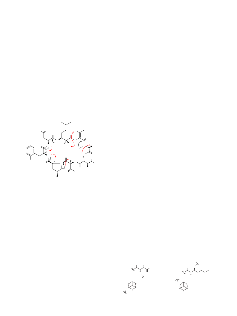
NOE NH/CH and NCH3/CH interactions and HMBC correlations
d 173.7 ppm.
a
a
acquired in CD3OH (Table 1 and Fig. 1).
The relative configuration of g-methylproline was determined
on the isolated amino acid residue obtained by acidic hydrolysis of
the parent peptide followed by HPLC separation.
Recently
a stereoselective access to all stereoisomers of
O
H
H2N
g
-methylproline residue has been published together with relevant
O
O
physical data.40 The two diastereoisomers can be differentiated on
the basis of 1H NMR data. Particular diagnostic are the chemical
shifts of H-3 diasterotopic methylene protons observed at dH 2.52
and 1.64 in the cis isomer and at dH 1.94 and 2.25–2.38 in the trans
isomer, and the coupling constant pattern of one of the H-5 protons
observed as a broad triplet (dH 2.87, J 10.6) in the cis isomer and as
double doublet (dH 2.83, J 11.5 and 8.6 Hz) in the trans isomer. The
O
N
H
N
H
H
HO
N
H
O
NH
N
O
H
HN
NH
O
H
O
H
N
H
H
OH
O
1H NMR spectral data of
perthamide C (see Experimental section) are consistent with the cis
stereochemistry.
g-methylproline isolated from parent
NH2
OH
O
MeO
The absolute configuration of the
determined by application of Marfey’s method. Marfey’s derivatives
of all stereoisomers of -methylproline has been prepared and
characterised, and it was found that they follow the usual elution
order, namely the -amino acid FDAA derivative is eluted from
a C-18 column before its corresponding
-isomer.40 Thus a small
sample of perthamide C (1) was hydrolysed and derivatised with
both enantiomers of Marfey’s reagent. By monitoring for FDAA-
methylproline at m/z 382, the 2S configuration was assigned,
g-methylproline residue was
Figure 1. Selected NOE (red) and HMBC (black) correlations for 1.
g
Long-range correlations between NH/NCH3 protons to carbonyl
carbons of adjacent amino acids allowed us to establish the fol-
L
lowing sequences: AHMHA-Asn-oTyr-
Moreover, connectivity of the MePro unit to ThrOMe residue was
indicated from an HMBC correlation between one of the MePro-
174.3 (C-1
gMePro and NMeGly-dAbu.
D
g
g
d-
g-
methylene protons (d 4.23) and the carbonyl at d
ThrOMe).
therefore we assign the cis-4-methyl-L-proline stereostucture to
Definitive confirmation of the sample structure was derived
from ESI MS/MS analysis. In addition to the pseudomolecular ion at
m/z 945.81 [MþH]þ, the ESI Q-TOF MS/MS spectrum provided
several fragment ion peaks. The detailed interpretation of the
fragmentation pattern confirmed both the sequence of amino acids
and the identity of the amidic-bearing amino acid residues. The
major peaks correspond to C-terminus fragments derived from
a homogeneous ring opening between the dehydrated threonine
(dAbu) and the N-methylglycine residue.
this subunit.
J-Based NMR configurational analysis method was used to de-
termine the relative configuration of -hydroxyasparagine residue
(Fig. 2). To support the assignment of an anti relative configuration
b
and to assign the absolute stereochemistry, all stereoisomers of
b
-hydroxyaspartate were prepared by ozonolysis of the corre-
sponding
-hydroxyphenylalanine stereoisomers.41 The so obtained
-hydroxyaspartic acids were used as standards in the Marfey’s
analysis, that evidenced the presence of (2R,3S)- OHAsn residue in
b
b
b
In particular, the signal at m/z 744.55 corresponding to the loss
of 201.26 amu from the pseudomolecular ion was indicative of the
presence of N-methylglycine and b-hydroxyaspartic acid residues.
Subsequently, the loss of 114.99 amu from the pseudo y6 ion was
attributed to the lack of ThrOMe (m/z at 629.56) residue, and the
further losses of 111.30 amu from pseudo y5 and 163.07 amu from
O
OH
O
HN
2
4
NH2
1
3
5
7
6
2
1
3
4
HN
O
OH
C1
C1
3JH2-H3 0.8 Hz (small)
3JH2-C4 1.4 Hz (small)
3JH3-C1 1.6 Hz (small)
2JH2-C3 -1.8 Hz (small)
3JH2-H3 1.0 Hz (small)
3JH2-C4 1.0 Hz (small)
2JH2-C3 -1.2 Hz (small)
2JH3-C2 -4.0 Hz (large)
pseudo y4 were indicative of the presence of
o-tyrosine.
Besides, several internal fragments have been detected; the
peak at 275.21 amu has been attributed to the dipeptide unit
methylproline/o-tyrosine; the fragment at 389.30 amu was
g
-methylproline and
HO
HN
C4
HN
HO
H3
H2
H2
H3
C4
g-
Figure 2. Relative configuration for AHMHA and bOHAsn residues in perthamide C (1).

 Festa, Carmen