Chemistry of Natural Compounds, Vol. 48, No. 5, November, 2012 [Russian original No. 5, September–October, 2012]
NEW CYCLOARTANE GLYCOSIDES FROM
Astragalus caucasicus AND A. galegiformis
*
M. D. Alaniya and T. I. Gigoshvili
UDC 547.918:547.926
In continuation of research on cycloartane triterpenoids from leaves of Astragalus caucasicus Pall. and stems of
A. galegiformis (Leguminosae L.), one compound from each was isolated and called D (1) and C (2) [2], respectively, in
13
addition to previously isolated compounds [1, 2] from enriched fractions. Based on IR, PMR, and C NMR spectral data,
these compounds were assigned to the cycloartane series [3] (Table 1).
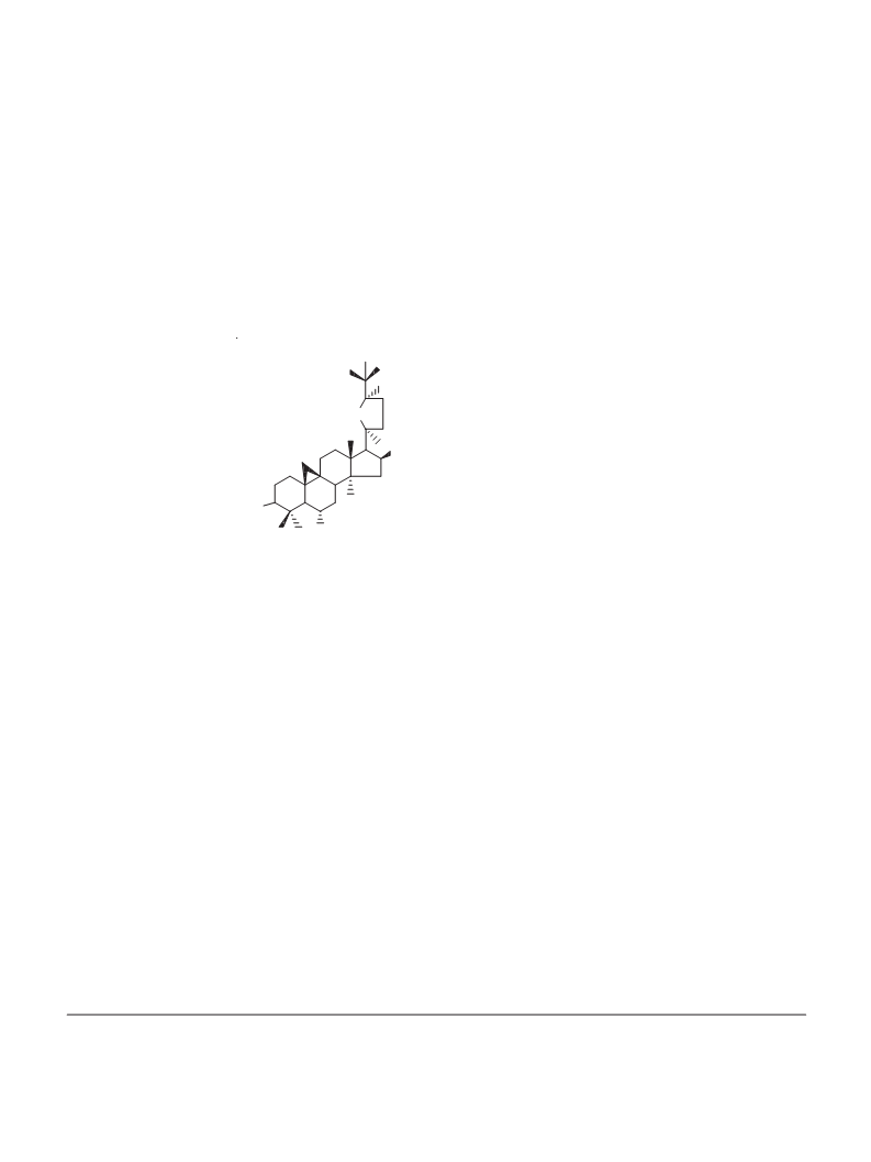
OH
H
O
OH
1: R = Glcp-(6ꢆ1)-Rhap, R = H
1
2: R = (2ꢃ-OAc)-Xylp, R = Xylp
RO
1
3: R = R = H
1
OR
1 - 3
1
Compound 1, white needle-like crystals, MW 798.3 g/mol (mass spectrometry), C H O , mp 220–222°C (MeOH).
42 70 14
–1
IR spectrum (KBr, ꢀ , cm ): 3600–3200 (OH), 3050 (cyclopropane ring CH ),
max
2
+
Acid hydrolysis of 1 and 2 produced a genin (3), 490 [M] , C H O , mp 194–196°C (MeOH). A solution of the
30 50
5
genin in acetone in the presence of H SO did not give an acetonide, indicating the absence of an ꢁ-diol group in the side
2
4
13
chain. The genin was identified by direct comparison with an authentic sample on TLC and C NMR data as cyclogalegigenin
[2]. Paper chromatography of the aqueous part of the hydrolyzate of 1 detected D-glucose and L-rhamnose; of 2, D-xylose.
Glycoside 1 was hydrolyzed by rhamnodiastase into cyclogalegigenin and rutinose [4].
13
According to a comparison of C NMR spectral data for 1 and 3, the carbohydrate unit in 1 was located on C-3 of the
genin (Table 1). The SSCC of the anomeric protons indicated that the monosaccharides had the pyranose form and the
ꢂ-configuration for D-glucose and the ꢁ-configuration for L-rhamnose [5].
Based on the analysis of the results, 1, which was isolated for the first time from A. caucasicus, was cyclogalegigenin
3-O-ꢂ-D-rutinoside, which we called cycloascauloside D.
+
Compound 2, white needle-like crystals, mp 184–188°C (aq. MeOH), [M] 796.25 (mass spectrometry), C H O .
42 68 14
–1
IR spectrum (KBr, ꢀ , cm ): 3600–3220 (OH), 3050 (cyclopropane ring CH ). The PMR spectrum showed at 2.01 ppm
max
2
a resonance for an acetyl (Table 1). Acid cleaved 2 on cyclogalegigenin and D-xylose. Base gave a less polar glycoside, acid
hydrolysis of which produced 3 and D-xylose.
Enzymatic hydrolysis of the glycoside by Helix plectotropis gave D-xylose and a monoside with mp 226–227°C
(CHCl :MeOH, 1:1), which was identified as cyclogaleginoside A [6].
3
A comparison of chemical shifts for Cꢃ and Cꢃꢃ atoms of 2 showed that C-1ꢃ underwent a diamagnetic shift by 2.69 ppm
[ꢄꢅ = ꢅ(C-1ꢃꢃ) 107.49 – ꢅ(Cꢃ) 104.80]. This indicated that the acyl group was located in D-xylose bonded to C-3 of the
aglycon. The strong-field shift of C-1ꢃ by 2.69 ppm indicated that the acetyl was located on the neighboring C atom (C-2ꢃ) [7].
I. G. Kutateladze Institute of Pharmaceutical Chemistry, Tbilisi, fax: (99532) 25 00 26, e-mail: merialania@yahoo.com.
Translated from Khimiya Prirodnykh Soedinenii, No. 5, September–October, 2012, pp. 809–810. Original article submitted
March 1, 2012.
©
914
0009-3130/12/4805-0914 2012 Springer Science+Business Media New York

 Alaniya