E.C. O'Neill et al. / Carbohydrate Research xxx (2017) 1e15
9
and glucose in the active site) will be the default reference structure
see Supplementary Fig. S10 for a structure-based sequence align-
ment). Like these enzymes, CDP contains a large -sandwich
domain that forms the majority of the dimer interface, connected
by a two -helix linker to an ( -barrel catalytic domain, and
ends with a small peripheral domain that adopts a two layered-jelly
roll fold (Fig. 7A and B, refer to Fig. 7B for domain colour-code).
Uniquely, CDP has a further ~120 amino acids at the N-terminus,
beginning with an extended arm that leads into a globular domain
by a sugar bound in the ꢁ1 subsite, and thus must be bound before
the glucan co-substrate in the phosphorolytic reaction, consistent
with it following a sequential Bi Bi mechanism. Beyond subsite þ1,
the acceptor site opens out and the glucan chain extends across a
wide U-shaped canyon formed at the dimer interface (Fig. 9A and
C), such that the residues occupying subsites þ2 and þ3 also
(
b
a
a/a)
6
interact with side-chains from the
b-sandwich domain of the
opposing subunit (Fig. 8). Although no electron density is present
for sugar residues beyond that occupying the þ3 subsite
(Supplementary Fig. S11), we cannot rule out the presence of the
two further residues expected for cellohexaose. Indeed, this may
indicate that the þ3 subsite delimits the extent of ordered binding
by the acceptor site. There are no large conformational changes
between the apo and ligand bound CDP structures (rms deviation of
0.692 Å for a dimer on dimer superposition), although a number of
side-chains become reoriented to engage with the substrates. In
particular, the carboxylate of the catalytic Asp624 is flipped about
comprised of a central, five-stranded, mixed
short -helices; the latter is unrelated to any structurally charac-
terised domain (Fig. 7). Both the domain and the N-terminal
arm interact with the -sandwich domain of the opposing subunit.
Indeed, a -strand ( 1) within the arm contributes to the one of the
sheets within the -sandwich (Fig. 7A, dashed red-circle, and C).
b-sheet, flanked by
a
a/b
b
b
b
b
Together, these additional interactions add substantially to the
2
dimer interfacial area: the total for CDP is ~4800 Å , as compared to
2
the value of ~3300 Å calculated for Cellovibrio gilvus CBP (PDB code
the C
a
-C
b
bond, to hydrogen bond with O3 of the ꢁ1 subsite sugar
3
1
QG0) using the PISA server [51]. Very recently, four structures of
/2-oligoglucan phosphorylase (SOGP) from Lachnoclostridium
b
-
and the oxygen of the scissile glycosidic linkage; additionally, the
adjacent residue within this “catalytic loop”, Cys625, also hydrogen
bonds to O2 of the þ1 subsite sugar (Fig. 8). Together, these in-
teractions cause the loop, which includes Trp622, to shift towards
phytofermentans were reported [52], which have slightly lower
DALI Z scores of ~32, with only 47% of the structure aligned to CDP
and, in contrast to all the aforementioned structures, they are all
monomeric. SOGP also differs from these enzymes in that it acts on
the bound substrate (Ca-Ca shift of 1.3 Å for Asp624). In general, the
majority of the interactions we observe with the ꢁ1 and þ1 subsite
sugars and the phosphate ion are structurally conserved in ligand
bound structures of CBP, but the correspondence is weaker for
SOGP (e.g. in PDB code 5H41), where the architecture necessarily
differs because the acceptor is bound in an orientation that is
orthogonal to that in the other enzymes [52]. Consistent with the
substrate preferences of CBP, its active site is significantly more
enclosed than that of CDP (Fig. 9B and D). This is largely due to three
structural features. Firstly, the catalytic loops differ in length, being
twelve residues longer in CDP (Fig. 9). Whilst the loops are struc-
turally similar up to and including the portion containing Asp624,
they adopt completely different conformations after Ile628. In the
case of one subunit of apo-CDP, the loop continues away from the
b
-1/2-glucan oligosaccharides rather than
saccharides. Like CDP, SOGP also has an extra, albeit much larger
~250 residues) N-terminal extension, again forming an extended
arm and a discrete domain, although the latter resembles the
b-1/4-glucan oligo-
(
b
-
sandwich domain common to all these phosphorylases, such that it
has two of these domains in tandem (Fig. 7). Remarkably, the
additional domain is placed relative to the remainder of the subunit
such that it aligns with the b-sandwich domain from the opposing
subunit of a superposed CDP/CBP-like dimer, thereby mimicking
the dimer interface of these latter enzymes. Moreover, the N-ter-
minal arm of SOGP interacts with the second
b-sandwich domain in
a similar way to the interaction seen between the N-terminal arm
of CDP and the
and C).
b-sandwich domain of the opposing subunit (Fig. 7A
active site forming a helix (
surface, before returning to the protein core; in the other subunit, a
short section following 17 is disordered. The latter is true for one
a17) that projects from the protein
a
2
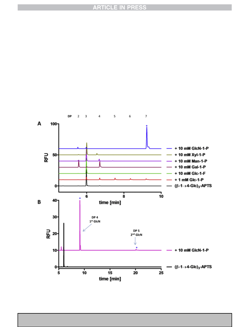
.7. Ligand bound structure of CDP
subunit in ligand-bound CDP (Fig. 9A and C), whereas in the other,
substantially more of this loop is disordered, with the exception of
the portion bearing Trp622 and Asp624. By contrast in CBP, the
catalytic loop is fully ordered, with the C-terminal portion folded
over the active site pocket (Fig. 9B and D). Secondly, an “adjacent”
loop, which packs against the catalytic loop, also impinges on the
active site cleft, but has a lesser impact in CDP as it is five residues
shorter than the equivalent loop in CBP. A final significant differ-
ence relates to how the acceptor pocket is defined by the opposing
The active site of CDP lies at one end of the (
catalytic domain and the substrate binding site is largely delineated
by the loops that connect the outer ring of -helices to the inner
ring. All attempts to co-crystallise CDP with a variety of substrates
resulted in poor quality crystals. However, when 10 mM cellohex-
aose was soaked into a crystal which had been grown in the
presence of 10 mM phosphate buffer, a dataset was collected to
6
a/a) -barrel of the
a
3
.0 Å resolution revealing additional electron density in the active
subunit, in particular, by two
a-helices (a6 and a7) within a loop of
site cleft, which was interpreted as a cellotetraose molecule with an
adjacent phosphate ion. Given the relatively low resolution of this
structure we were unable to be certain of the conformation of the
the -sandwich domain. In the more open CDP pocket, two side-
b
chains from this “opposing loop” contribute to the acceptor bind-
ing site: Asp297 hydrogen bonds to O3 of the þ2 subsite sugar, and
Tyr300 forms a stacking interaction with it. There is also a further
hydrogen bond to O1 of the þ3 subsite sugar, via the side-chain of
Glu328 in a different loop of the opposing subunit (Fig. 8 and 9,
Supplementary Fig. S11). Compared to CDP, the juxtaposition of the
two subunits differs in CBP due to a global rotation about an axis
that is perpendicular to, and almost bisects, the two-fold axis
relating one half of the dimer to the other (Fig. 7C). In the Cellovibrio
sugar rings: we therefore chose to treat them all as
b
-1/4 linked
4
C
1
chairs, which gave a reasonable fit to the electron density with
good geometrical parameters. The glucan was oriented such that
the non-reducing terminal glycosidic bond was located between
the phosphate and Asp624, the expected general acid catalyst
(Fig. 8 and Supplementary Fig. S11). Thus, the glucan spanned
subsites ꢁ1 to þ3 of the active site pocket. The donor site is
completely buried and does not extend beyond the ꢁ1 donor
subsite where it is terminated by the side-chain of Trp622, the so-
called “hydrophobic platform” residue that is structurally
conserved in the close homologues. This is important to exclude
water around the region of the scissile bond. The phosphate is
located in an adjacent lobe of the active site cleft that is closed off
ꢀ
gilvus CBP (PDB code 3QG0) the rotation is ~16 (although it is
similar for others), and this has the effect of significantly narrowing
the canyon between the two subunits and, thereby driving the
opposing loop towards the active site (Fig. 9B and D). This fore-
shortens the acceptor binding pocket, such that in CBP, Gln165 in
the equivalent of a7 in the opposing loop, hydrogen bonds to O5 of
Please cite this article in press as: E.C. O'Neill, et al., Cellodextrin phosphorylase from Ruminiclostridium thermocellum: X-ray crystal structure

 O'Neill, Ellis C.