Silver Nanoparticles and Chloroaurate Ions
J. Phys. Chem. B, Vol. 109, No. 42, 2005 19625
the film was negligible (data not shown for brevity) and nowhere
comparable to that obtained in the case of transmetalation in
the presence of the ODA monolayer (Figure 3).
ions and the effusion of Ag+ ions during the transmetalation
reaction thereby limiting the reaction to the stage where only a
thin shell of gold is formed around the silver core. It is known
that a gold shell of 3-5 atomic layers is sufficient to dampen
the surface plasmon vibrations of the underlying silver core
completely2b,c and could thus explain the UV-vis spectroscopy
trends observed.
In conclusion, we have shown that the facile transmetala-
tion reaction between hydrophobic silver nanoparticles and
aqueous chloroaurate ions can be carried out at the air-water
interface. Such reactions with nanoparticles assembled into
close-packed structures at interfaces could lead to metallic cross-
linking of the nanoparticles and synthesis of extended nano-
particulate structures as shown. In the presence of suitable
electrostatic barriers, the assembly of the nanoparticles after
transmetalation reaction leads to interesting honeycomb-like
superstructures. The possibility of connecting nanoparticles in
close proximity via electrical contacts shows potential for
generation of conducting electrodes with nanoparticles as
building blocks and in the design of optical coatings and
chemical/biological sensors.
It would be of interest to study the transmetalation reaction
between LB films of Ag-ODA nanoparticles during immersion
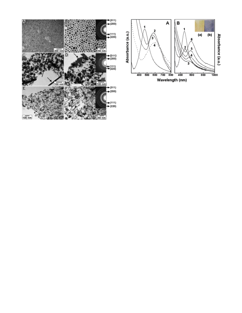
in HAuCl4 solution. The UV-vis absorption spectra recorded
from a 20 ML LB film of Ag-ODA nanoparticles measured
as a function of time of immersion in 10-3 M HAuCl4 solution
is shown in Figure 2B. It is observed that as the transmetalation
reaction proceeds, there is a progressive decrease in the intensity
of the silver plasmon band at 470 nm (curve 2, 10 min of
reaction) that is then followed by the appearance of a separate
and distinct absorption band from metallic gold nanoparticles
at ca. 560 nm after 1 h of reaction. Further immersion in the
chloroaurate ion solution leads to an increase in the gold
plasmon band intensity and a small shift in the peak position to
570 nm (curves 4-6). The reaction is complete after nearly 48
h of reaction and is much slower than the transmetalation
reaction between the silver nanoparticles and gold ions at the
air-water interface. We believe that this difference in reaction
rates is due to the fact that the infusion rate of gold ions and
effusion of Ag+ ions arising from the transmetalation reaction
would be much smaller in the case of the LB films of the
hydrophobic silver nanoparticles during immersion in chloro-
auric acid solution. Such a constraint does not occur at the air-
water interface where the silver nanoparticles fully access the
subphase gold ions with no barrier to metal ion diffusion. The
color of this film turned from yellow (inset of Figure 2B, slide
“a”) to a deep blue upon completion of the reaction with gold
ions (inset of Figure 2B, slide “b”). The blue color indicates
the interparticle surface plasmon coupling in a monolayer of
gold nanoparticles.15 The 20 ML Ag-ODA LB film after 48 h
of immersion in chloroauric acid solution was dissolved in
chloroform, and the nanoparticles were imaged by TEM (parts
C and D of Figure 5). It is observed that the particles are well
separated from one another and that the particles are bigger
(average size ∼50 nm) than the as-prepared silver nanoparticles
(20 ( 6 nm). This may be attributed to the fact that the AgCl
formed after the transmetalation reaction is not able to leach
out fully due to entrapment in the lipid monolayer and stays in
the form of solid AgCl. We do not observe any interconnection
between the nanoparticles possibly because during immersion
of the Ag-ODA LB film in the chloroauric acid solution, the
film swells and leads to considerable separation between the
silver nanoparticles. Transmetalation thereafter may not be
capable of interlinking the nanoparticles, and they merely grow
in size. We note that the UV-vis absorption curves recorded
from the LB films after transmetalation reaction (parts A and
B of Figure 2) show the absorbance at ca. 580 nm, indicative
of the formation of gold nanostructures. Usually it is observed
that the transmetalation reaction between silver nanoparticles
and gold ions in either aqueous13,14 or organic18 medium leads
to the formation of hollow gold nanostructures. However, in
the present study, the UV-vis spectroscopy results (parts A
and B of Figure 2) indicate the formation of either pure solid
gold nanostructures or bimetallic nanostructures (gold-coated
silver cores).19 TEM results support the UV-vis spectroscopy
data showing the presence of both solid gold (parts D and F of
Figure 1 and Figure 4F) and bimetallic Au-Ag (Figure 4F,
particles indicated by arrows) nanostructures. While the exact
reasons for this difference are not understood at this moment,
we believe the ODA-capping layer surrounding the sacrificial
silver nanoparticles may be playing an important role. The ODA
Acknowledgment. A.S. thanks the Council of Scientific and
Industrial Research, Government of India, for a research
fellowship during the initial stages of this work.
References and Notes
(1) Sastry, M. In Colloids and Colloid Assemblies: Synthesis, Modi-
fication, Organization and Utilization of Colloid Particles; Caruso, F., Ed.;
Wiley-VCH: Berlin, 2003; Chapter 12, p 369. (b) Sastry, M. In Handbook
of Surfaces and Interfaces of Materials; Nanostructured Materials, Micelles
and Colloids; Nalwa, H. S., Ed.; Academic Press: London, 2001; Vol. 3,
Chapter 2, p 87. (c) Fendler, J. H. Nanoparticles and Nanostructured
Films: Preparation, Characterization and Applications; Wiley-VCH:
Weinheim, Germany, 1998. (d) Rotello, V. Nanoparticles: Building Blocks
for Nanotechnology; Kluwer Academic/Plenum Publishers: New York,
2004. (e) Horiuchi, S.; Sarwar, M. I.; Nakao, Y. Adv. Mater. 2000, 12,
1507. (f) Horiuchi, S.; Fujita, T.; Hayakawa, T.; Nakao, Y. Langmuir 2003,
7, 2963.
(2) El-sayed, M. A. Acc. Chem. Res. 2001 34, 257. (b) Henglein, A. J.
Phys. Chem. 1993, 97, 5457. (c) Henglein, A. Chem. Rev. 1989, 89, 1861.
(d) Alivisatos, A. P. J. Phys. Chem. 1996, 100, 13226. (e) Schmid, G.
Clusters and Colloids: From Theory to Application; VCH: Weinheim,
Germany, 1994. (f) Perenboom, J. A. A. J.; Wyder, P.; Meier, P. Phys.
Rep. 1981, 78, 173. (g) Hughes, A. E.; Jain, S. C. Adv. Phys. 1979, 28,
717.
(3) Andres, R. P.; Bein, T.; Dorogi, M.; Feng, S.; Henderson, J. I.;
Kubiak, C. P.; Mahoney, W.; Osifchin, R. G.; Reifenberger, R. Science
1996, 272, 1323. (b) Kim, S. H.; Medeiros-Ribeiro, G.; Ohlberg, D. A. A.;
Williams, R. S.; Heath, J. R. J. Phys. Chem. B 1999, 103, 10341. (c) Beverly,
K. C.; Sampaio, J. F.; Heath, J. R. J. Phys. Chem. B 2002, 106, 2131. (d)
Kim, B.; Tripp, S. L.; Wei, A. J. Am. Chem. Soc. 2001, 123, 7955.
(4) Xia, D.; Biswas, A.; LI, D.; Brueck, S. R. J. AdV. Mater. 2004, 16,
1427. (b) Xia, D.; Brueck, S. R. J. Nano Lett. 2004, 4, 1295.
(5) Lee, K.; Pan, F.; Carroll, G. T.; Turro, N. J.; Koberstein, J. T.
Langmuir 2004, 20, 1812. (b) Horiuchi, S.; Fujita, T.; Hayakawa, T.; Nakao,
Y. AdV. Mater. 2003, 15, 1449.
(6) Xia, Y.; Whitesides, G. M. Angew. Chem., Int. Ed. 1998, 37, 550.
(b) Gorman, C. B.; Beibuyck, H. A.; Whitesides, G. M. Chem. Mater. 1995,
7, 526. (c) Zhao, X. M.; Xia, Y.; Whitesides, G. M. J. Mater. Chem. 1997,
7, 1069.
(7) Santhanam V.; Andres, R. P. Nano Lett. 2004, 4, 41.
(8) Sastry, M.; Patil, V.; Mayya, K. S.; Paranjape, D. V.; Singh, P.;
Sainkar, S. R. Thin Solid Films 1998, 324, 239. (b) Perez, H.; Lisboa de
Sousa, R. M.; Pradeau, J. P.; Albouy, P. A. Chem. Mater. 2001, 13, 1512.
(c) Swami, A.; Kumar, A.; Selvakannan, PR.; Mandal, S.; Sastry, M. J.
Colloid Interface Sci. 2003, 260, 367. (d) Selvakannan, PR.; Swami, A.;
Srisathiyanarayanan, D.; Shirude, P. S.; Pasricha, R.; Mandale, A. B.; Sastry,
M. Langmuir 2004, 20, 7829. (e) Wang, W.; Chen, X.; Efrima, S. J. Phys.
Chem. B 1999, 103, 7238. (f) Heath, J. R.; Knobler, C. M.; Leff, D. V. J.
Phys. Chem. B 1997, 101, 189. (g) Burghard, M.; Philipp, G.; Roth, S.;
von Klitzing, K.; Pugin, R.; Schmid, G. AdV. Mater. 1998, 10, 842. (h)
Bourgoin, J. F.; Kergueris, C.; Lefevre, E.; Palacin, S. Thin Solid Films
1998, 327-329, 515. (i) Chen, X. Y.; Li, J. R.; Jiang, L. Nanotechnology
-
monolayer could act as a barrier toward the infusion of AuCl4

 Pasricha, Renu