S. Hussain et al. / Polyhedron 117 (2016) 64–72
67
The 13C NMR spectrum of 3 displayed the expected signals of its
ligand skeleton and the coordinated organotin moieties. The
upfield shift of the resonance signal for the carboxylate carbon
atom of the free ligand (Na2LꢀH2O) from d 179.3 to 171.1–
172.7 ppm in complexes 1–3 can be attributed to the carboxy-
late-tin linkage and indicates that the carbon atom of carboxylate
ion bound to the tin center is more electron rich compared to that
of three methyl radicals and complete decarboxylation to generate
the [C7H12NO4Sn2]+ (m/z = 414 (37%)) and [C8H21NSn2]+ (m/z = 371
(10%)) ions, respectively. In the negative mode, the appearance of a
peak with m/z = 457 (6%) corresponds to the formation of the [C10
H
-
19NO4Sn2]ꢁ ion. The EIMS of complex 2 did not display a molec-
ular ion peak. It evolved a triphenyltin radical to generate the
[C22H20NO4Sn]+ (m/z = 482 (16%)) ion, which further loses a phenyl
radical in each successive step to yield the [C16H15NO4Sn]+ 405
(100%), [C10H10NO4Sn]+ 328 (31%) and [C4H5NO4Sn]+ 251 (35%)
ions step by step. The parent species 2 may also split into a triph-
0
bound with sodium. Also the methylene carbon atoms (C-3,3 ) in
the products 1 and 2 are more shielded (d 51.2–51.3 ppm) as com-
pared to those in the ligand precursor (d 51.9 ppm). However, the –
CH2– signal did not show any shift in complex 3 as compared to the
ligand; the appearance of this peak at the same position in the
ligand and product 3 has also been verified by 1H NMR spec-
troscopy. The carbon atoms of the methyltin(IV) and dichlorodi-
methyltin(IV) moieties in complex 3 exhibited similar nuclear
magnetic resonance behavior in solution, which is demonstrated
by the appearance of their 13C NMR chemical shifts at the same
position (d 23.5 ppm). A penta-coordinated environment around
the metal center in the triphenyltin(IV) derivative 2 was verified
through the appearance of resonance shifts at d 142.7 ppm for
the ipso-carbon atom. It is known that the chemical shifts d(13C)ipso
of the phenyl groups in the SnPh3 moiety lie in the range d 135.9–
138.7 ppm, corresponding to the value shown by tetrahedrally
coordinated Sn atom. For penta-coordinated triphenyltin(IV) car-
boxylate complexes d(13C)ipso will be observed at approximately
4 ppm higher [24].
enyltin cation (m/z = 351(24%)) whose further degradation contin-
+
ues until the formation of the Sn cation. The [C12H10
]
fragment
ion, m/z = 154 (86%), corresponds to the formation of a biphenyl
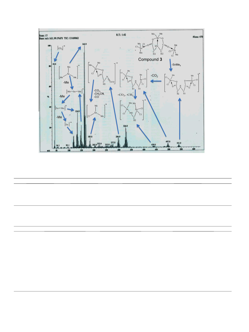
cation. Fig. 1 shows the EI-mass spectrum and the structures of
important fragment ions of coordination compound 3. The mole-
cules of 3 start to decompose through the expulsion of tetram-
ethyltin(IV) (SnMe4) to yield the [C6H11Cl4NO4Sn2]+ ion (m/
z = 541 (4%)), which first loses CO2 and then CO2 and CH2 radicals
to leave behind the deposits of the [C5H11Cl4NO2Sn2]+ (m/z = 497
(7%)) and [C3H9Cl4NSn2]+ (m/z = 439 (3%)) ions, respectively. The
dimethyltin cation (m/z = 150 (21%), may lose one or both sub-
stituents to yield [CH3Sn]+ (m/z = 135 (37%)) and [Sn]+ (m/z = 120
(13%)), respectively or it may combine with a methyl radical to
yield [C3H9Sn]+ (m/z = 165 (100%)). The peak at m/z = 185 (9%) cor-
responds to the chlorodimethyltin(IV) cation and explores the exis-
tence of a chloro-substituted tin(IV) compound, which has also
been confirmed by single crysal XRD analysis of 3.
Thermogravimetric analyses (TGA) of the ligand (Na2LꢀH2O) and
complex 2 were performed under an N2 atmosphere to compare
their degradation patterns, thermal stabilities and percentage puri-
ties. The total weight loss was theoretically calculated and com-
pared with the experimental results. The thermally decomposed
data agreed well with the expected chemical composition of the
ligand and the product complex. The data showed that while the
ligand (Na2LꢀH2O) is decomposed up to 794.12 °C, leaving behind
a final residual mass (end product) equivalent to Na2CO3, complex
2 undergoes thermal degradation up to a slightly higher tempera-
ture (800.00 °C) to evolve volatile molecules/radicals of (4Phꢀ,
SnH4, 2CO, CCHꢀ, Nꢀ) and depositing a final residue of SnO2. The
results are thus completely consistent with the molecular compo-
sition of product 2. The thermal decomposition data are summa-
rized in Table 3.
3.4. Mass spectrometry and thermogravimetric analyses
The electron ionization mass spectrum (EI-MS) was recorded for
complex 1. Product 2 was investigated by electrospray ionization
mass spectrometry (ESI-MS). The mass fragmentation data have
been summarized in Table 2. The ions containing the tin(IV) ion
appeared in the spectra as a series of peaks close to each other
due to isotopic effects. The relative intensities reported here were
qualitatively estimated from the ion current for the 120Sn peak in
each species, and must be regarded as approximate. The ESI-MS
(positive mode) of 1 shows the appearance of an M-20 peak (m/
z = 459 (19%)) corresponding to the loss of water and a hydrogen
molecule from the parent species. The evolution of H2 generates
a double bond between the imino nitrogen and methylene carbon
atoms in the ligand skeleton of the complex. Further fragmentation
of [C10H21NO4Sn2]+ (m/z = 459 (19%)) is continued by the removal
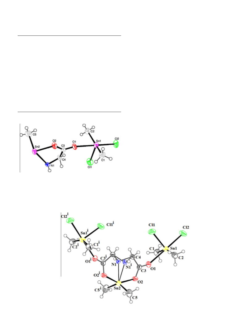
3.5. XRD crystallography
Table 4 represents the important crystallographic data of com-
pound 3; selected bond angles and bond lengths are given in
Table 5. The single crystal XRD study of 3 has proven it to be a trin-
uclear complex and one of the very few reported examples of
organotin(IV) carboxylates showing the simultaneous incorpora-
tion of dimethyltin(IV) and dichlorodimethyltin(IV) moieties.
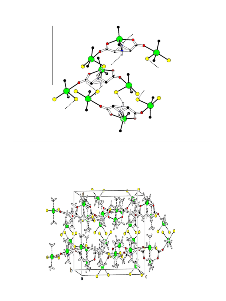
There is evidence of intermolecular N–Hꢀ ꢀ ꢀCl interactions in the
crystal lattice (Figs. 4 and 5). Each discrete molecule (Figs. 2 and
3) consists of two kinds of tin center, i.e. Sn1 in each of the two
dichlorodimethyltin(IV) fragments and Sn2 in a dimethyltin(IV)
moiety. The ligand reacts with the metal in a pentadentate fashion,
contributing all of its four carboxylate oxygen atoms and the imino
nitrogen in coordination. A structural comparison of coordination
compound 3 with the reported complexes [Me2Sn(L)(MeOH)]2,
Table 2
Mass spectral data of complexes 1–3.
Comp.
No.
MS, m/z (%)
1
Positive mode: [C10H25NO5Sn2]+ 479 (n.o)#, [C10H21NO4Sn2]+ 459
(19), [C7H12NO4Sn2]+ 414 (37), [C8H21NSn2]+ 371 (10),
[C6H16NSn2]+ 342 (40), [C5H12NSn2]+ 326 (100), [C7H12NO4Sn]+ 294
(50), [C5H9NO2Sn]+ 236 (18), [C4H6NO2Sn]+ 220 (1).
Negative mode: [C10H25NO5Sn2]ꢁ 479 (n.o)#, [C10H19NO4Sn2]ꢁ 457
(6), [C9H19NO2Sn2]ꢁ 413 (12), [C8H19NSn2]ꢁ 369 (10),
[C6H15NSn2]ꢁ 341 (2), [C5H12NSn2]ꢁ 326 (2), [C2H6NSn2]ꢁ 284 (12),
[C6H10NO4Sn]ꢁ 280 (100), [C4H8NO2Sn]ꢁ 222 (9).
2
3
[C40H35NO4Sn2]+ 833 (n.o)#, [C22H20NO4Sn]+ 482 (16),
[C16H15NO4Sn]+ 405 (100), [C18H15Sn]+ 351 (24), [C10H10NO4Sn]+
328 (31), [C12H10Sn]+ 274 (16), [C4H5NO4Sn]+ 251 (35), [C6H5Sn
197]+ (14), [C12H10
(7).
]
+ 154 (86), [Sn]+ 120, [C6H5]+ 77 (12), [C4H3]+ 51
[Bu2Sn(L)(H2O)],
[Ph2Sn(L)(H2O)]
and
[Bu2Sn(PhL)(H2O)]
[19,20,22] of the same ligand indicates that the ligand ion (Lꢁ2
)
[C10H23Cl4NO4Sn3]+ 721 (n.o)#, [C6H11Cl4NO4Sn2]+ 541 (4),
[C5H11Cl4NO2Sn2]+ 497 (7), [C3H9Cl4NSn2]+ 439 (3),
[C6H8ClNO3Sn]+ 297 (11), [C2H6SnCl]+ 185 (9), [C3H9Sn]+ 165 (100),
[C2H6Sn]+ 150 (21), [CH3Sn]+ 135 (37), [Sn]+ 120 (13), [CO2]+ 44
(82).
acts in various multidentate modes and smaller molecules like
MeOH or H2O are also involved in coordination with the tin(IV)
ions. However, the synthesis of the investigated complex 3 does
not involve the incorporation of either water or methanol mole-
cules in its crystal lattice.
#
Molecular ion peak (M+); n.o. = not observed.

 Hussain, Shabbir