A R T I C L E S
Metallo et al.
water again and then stored frozen at -20 °C. While there is no
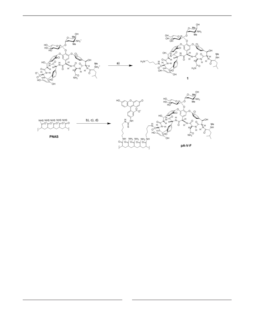
indication (from NMR of pA-V-F) that reaction between the nitrogen
of the hindered N-terminus or of the vancosamine moiety of 1 and
PNAS occurs, a low level of multipoint attachment to the polymer
backbone that would result from reaction at these additional sites cannot
be definitely excluded.
to proceed for 30 min at room temperature. The chips were removed
from the solution, washed with distilled deionized water and with
ethanol, and dried under a stream of nitrogen.
Surface Plasmon Resonance Spectroscopy. All SPR experiments
were carried out on a BIAcore 1000 instrument. Our substrates were
glued into the commercial BIAcore cartridges (after removing the
manufacturer’s substrate) and then docked into the BIAcore instrument.
PBS (10 mM phosphate, 138 mM NaCl, and 2.7 mM KCl, pH 7.4)
was prepared in distilled deionized water. Solutions of IgGF, pA, pA-
V-F, and pA-F were prepared in PBS.
Ellipsometry. Ellipsometric measurements were carried out on a
commercial ellipsometer (AutoEL II, Rudolph, Inc.). Substrates for
ellipsometry were prepared by evaporating titanium (1.5 nm, as an
adhesion layer) and then gold (100 nm) onto silicon wafers. We
determined the optical constants for each substrate. The substrates were
then immersed in ethanolic solutions of a mixture of HS(CH2)11(OCH2-
CH2)3OH (1.8 mM) and HS(CH2)11(OCH2CH2)6OCH2COOH (0.2 mM)
overnight. The coupling of N-R-Ac-KDADA to the mixed SAMs was
carried out as described above. The SAMs presenting DADA were
immersed in a solution of pA-V-F (10 µM vancomycin, 2 µM
fluorescein) in PBS for 6 h. The substrate was rinsed with PBS and
then with distilled water and dried under a stream of nitrogen.
Ellipsometric thicknesses were determined (at each step) using the
previously determined optical constants.
Characterization of Polymers. Poly(N-acryloyloxysuccinimide) was
hydrolyzed by a reaction with aqueous sodium hydroxide (0.1 N). The
polymer was dialyzed against deionized water, followed by lyophiliza-
tion, and the molecular weight distribution was characterized by GPC:
Mw ) 96 500; Mn ) 65 000; PDI ) 1.48; degree of polymerization
≈900. The fractional coupling of fluorescein and vancomycin on the
1
polymers was determined by H NMR and absorption spectroscopy.
Characteristic aromatic peaks of fluorescein were integrated and
compared with the integrated signal from the protons on the polyacryl-
amide backbone to determine the percent substitution with fluorescein.
The NMR signals from vancomycin on pA-V-F were too broad to
provide reliable integrated values, so the percent substitution was
determined based on the vancomycin-to-fluorescein ratio. Due to the
method of synthesis, the mole percent substitution of fluorescein on
the pA-V-F polymer was the same as on the pA-F polymer. The mole
percent vancomycin on pA-V-F was determined using its absorbance
at 280 nm, where both vancomycin and fluorescein absorb, and 450
nm, where only fluorescein absorbs. The molar absorptivity at 280 and
450 nm was determined from Beer’s law plots of vancomycin (ꢀ280
)
Polarized Infrared External Reflectance Spectroscopy. PIERS
spectra were measured using a DigiLab Fourier transform infrared
spectrometer (BioRad) equipped with a liquid N2-cooled mercury-
cadmium-telluride detector. The p-polarized light was incident at 80°
relative to the surface normal of the substrate. Substrates were prepared
in a manner identical to those used for ellipsometric measurements.
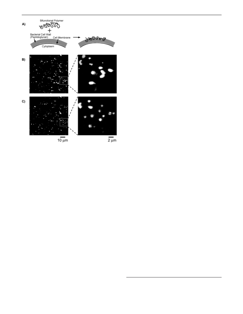
Bacterial Fluorescence. Bacteria (E. faecalis, ATCC 49332) were
grown overnight in brain-heart infusion broth at 37 °C, with shaking.
The bacteria were harvested by centrifugation and washed by resus-
pension in PBS and centrifugation. The bacteria were then resuspended
to an OD600 ) 1 in PBS containing 5 µM SYTO 63 (Molecular Probes)
and pA-V-F (15 µM in vancomycin, 3 µM in fluorescein). After 30
min at room temperature, 50 µL of the bacterial suspension was
transferred to an ELF spin filter (0.2-µm pore size, Molecular Probes)
and centrifuged to remove the polymer- and dye-containing solution.
The bacteria were washed by being resuspended in 200 µL of PBS
and centrifuged. Samples were washed three times. After the final
washing, the bacteria were resuspended in a one-to-one mixture of PBS
and Slowfade antifade reagent (Molecular Probes). Control samples
using SYTO 63 and pA-F, or only SYTO 63 were prepared using the
same procedure. Samples containing the Gram-negative bacteria E. coli
were prepared in the same manner as the E. faecalis samples. Bacterial
samples were imaged using a Leica confocal microscope equipped with
an Ar-Kr laser.
6030 M-1 cm-1; ꢀ450 ) 0 M-1 cm-1) and fluorescein cadaverine (ꢀ280
) 20 800 M-1 cm-1; ꢀ450 ) 20 600 M-1 cm-1). From these values and
the absorbance of pA-V-F at the two wavelengths, the concentration
of vancomycin and fluorescein was determined. The ratio of their
concentrations, in conjunction with the mole percent substitution of
fluorescein on the polymer, yielded the mole percent vancomycin. The
polymers used in this work (pA, pA-V-F, pA-F) are soluble in water
and PBS.
Preparation of Gold Substrates Presenting Mixed SAMs. Gold
substrates were prepared by evaporating titanium (1.5 nm, as an
adhesion layer) and then gold (38 nm) onto glass cover slips (0.2 mm
thick, No. 2, Corning).30 Gold substrates were immersed in ethanolic
solutions of a mixture of HS(CH2)11(OCH2CH2)3OH (1.8 mM) and HS-
(CH2)11(OCH2CH2)6OCH2COOH (0.2 mM) overnight.30
Coupling Ligands to Carboxylic Acid Groups in Mixed SAMs.
The tripeptide N-R-Ac-KDADA was coupled to mixed SAMs by amide
bond formation between the ꢀ-amino group of lysine and the carboxylic
groups on the surface of mixed SAMs. The procedure was described
previously by Lahiri et al.30 Briefly, the SAM-coated gold chips were
immersed in a solution of EDC (0.1 M) and NHS (0.4 M) in deionized
water for 7 min at room temperature; this treatment converted the
carboxylic acid groups into NHS active esters. The chips were removed
from the solution of EDC and NHS and immersed without rinsing in
a phosphate-buffered (25 mM; pH 8.1) aqueous solution of the tripeptide
(2 mg/mL); the coupling reaction was allowed to proceed for 30 min
at room temperature. The chips were removed from the solution of
peptide, washed with distilled, deionized water, and with ethanol, and
dried under a stream of nitrogen. Fluorescein cadaverine was coupled
to the mixed SAMs using a similar procedure. Briefly, after activating
the carboxylic acid groups of the mixed SAMs as NHS esters, the chips
were dipped into a solution of fluorescein cadaverine (2 mg/mL) and
triethylamine (10 µL/mL) in DMF. The coupling reaction was allowed
Acknowledgment. This research was supported by the
National Institutes of Health (NIH GM 30367), DARPA, and
Space and Naval Warfare Systems Center, San Diego. S.J.M.
and R.E.H. acknowledge postdoctoral fellowships from the NIH.
We thank Professor Paul Laibinis (MIT) for use of the FTIR
instrument in his laboratory.
JA030045A
9
4540 J. AM. CHEM. SOC. VOL. 125, NO. 15, 2003

 Metallo, Steven J.